. 2020 Dec 5;10(23):e3842.
doi: 10.21769/BioProtoc.3842.

Analysis of B Cell Migration by Intravital Microscopy

Affiliations

Analysis of B Cell Migration by Intravital Microscopy

Michael Schnoor et al. Bio Protoc. .

Abstract

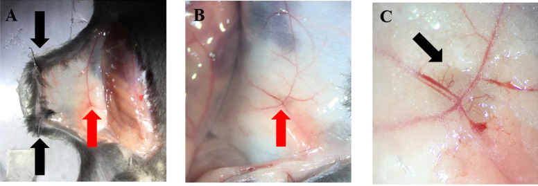
During immune responses, B cells home to lymph nodes (LNs), where they encounter antigens. Homing starts with capture and L-selectin-dependent rolling on the activated endothelium of high endothelial venules (HEV). After recognition of chemokines presented on HEV, activation of B cell integrins occurs mediating firm arrest. Subsequently, B cells crawl to the spot of extravasation to enter the LN. Extravasation can be visualized and quantified in vivo by intravital microscopy (IVM) of the inguinal LN. Here, we describe an established protocol that permits detailed in vivo analysis of B cell recruitment to LN under sterile inflammatory conditions. We describe data acquisition, exportation, quantification, and statistical analysis using specialized software. IVM of LN is a powerful technique that can provide a better understanding of B cell migratory behavior during inflammation in vivo.

Keywords: B cells; Extravasation; Intravital microscopy; Lymph node; Migration; Rolling.


Conflict of interest statement

Competing interestsThe authors declare no competing or financial interests.

Figures

Figure 1.
Figure 1.. Schematic representation of the localization of relevant organs involved in the IVM of the LN.
The trachea is shown here as a reference for the localization of the carotid artery, which is used for posterior catheterization. The localization of the spleen is shown for its resection in a donor mouse to isolate B cells. The two blue arrows in the lower part of the figure point to the right and left inguinal LN which are locally activated by injection of CXCL12 close to it. Both LNs can be used for IVM.
Figure 2.
Figure 2.. B cell enrichment and stimulation.
A. After B cell enrichment, purity was assessed by flow cytometry employing a monoclonal antibody against B220 coupled to AF488. Typically, a purity over 90% is obtained using α-Thy1 antibody-coated Petri dishes. B. CD44 expression on B cells increases after stimulation with LPS and IL-4 for 48 h.
Figure 3.
Figure 3.. Carotid artery catheterization.
A. A hairpin-like polyester thread is drawn below the carotid artery: one thread is placed towards the thorax; the other one towards the head. B. Close the blood flow towards the brain with a double knot (red arrow); with the other thread, make a loose knot (black arrow). C. Place a straight clamp to close the artery. D. Catheterize the carotid artery; fix the catheter with two knots and release the clamp from the artery. Blood will invade the inner part of the catheter (black arrow). The double arrow indicates the orientation of the mouse.
Figure 4.
Figure 4.. Microscope set-up, superfusion system and customized animal stage.
The superfusion system is composed of a hot stirring plate set at 45 °C to warm the superfusion buffer (PBS or 0.9% saline). With this setting, the buffer reaches the tissue at around 37 °C (this needs to be optimized in each laboratory). The buffer flask with an exit at the bottom is connected to a plastic tube that is fixed with regular tape to the 20x objective of the microscope. The customized animal stage is 25 cm long and 15 cm wide. This animal stage has the purpose of controlling the flow of PBS used to moisturize the exposed tissue. Additionally, it contains a little ring filled with silicon where the retracted skin can be fixed with needles (red arrow). This stage can be used for IVM of the LN, the cremaster muscle, and the mesentery. In the lower right part of the figure, an example of how the mouse is fixed to the animal stage is shown. Also, it exemplifies how the catheter should be fixed and how the mouse preparation should look prior IVM).
Figure 5.
Figure 5.. LN preparation.
A and B. Secure the abdominal skin to the animal stage with two pins (black arrows); the inguinal LN should be visible (red arrows). C. Appearance of the LN after removal of surrounding connective and adipose tissue. HEV are clearly visible using a dissection microscope (black arrow).
Figure 6.
Figure 6.. Identification of HEVs.
The number IV refers to the thinnest HEV, while the number I denotes the thickest HEV in the inguinal LN. The labeled B cells can be observed interacting with the HEV. (Objective 20x) Scale bar 50 µm.

References

    1. Andorko J. I., Tostanoski L. H., Solano E., Mukhamedova M. and Jewell C. M.(2014). Intra-lymph node injection of biodegradable polymer particles. J Vis Exp(83): e50984. - PMC - PubMed
    1. Chayen A. and Parkhouse R. M.(1982). B cell subpopulations in the mouse: analysis with monoclonal antibodies NIM-R2 and NIM-R3. Eur J Immunol 12(9): 725-732. - PubMed
    1. De Pascalis C. and Etienne-Manneville S.(2017). Single and collective cell migration: the mechanics of adhesions. Mol Biol Cell 28(14): 1833-1846. - PMC - PubMed
    1. Gavins F. N. and Chatterjee B. E.(2004). Intravital microscopy for the study of mouse microcirculation in anti-inflammatory drug research: focus on the mesentery and cremaster preparations. J Pharmacol Toxicol Methods 49(1): 1-14. - PubMed
    1. Giron-Perez D. A., Vadillo E., Schnoor M. and Santos-Argumedo L.(2020). Myo1e modulates the recruitment of activated B cells to inguinal lymph nodes. J Cell Sci 133(5). - PubMed

LinkOut - more resources