. 2020 Dec 3;6(6):e534.
doi: 10.1212/NXG.0000000000000534. eCollection 2020 Dec.

New recessive mutations in SYT2 causing severe presynaptic congenital myasthenic syndromes

Affiliations

New recessive mutations in SYT2 causing severe presynaptic congenital myasthenic syndromes

Stéphanie Bauché et al. Neurol Genet. .

Abstract

Objective: To report the identification of 2 new homozygous recessive mutations in the synaptotagmin 2 (SYT2) gene as the genetic cause of severe and early presynaptic forms of congenital myasthenic syndromes (CMSs).

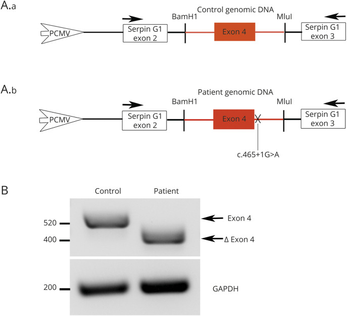
Methods: Next-generation sequencing identified new homozygous intronic and frameshift mutations in the SYT2 gene as a likely cause of presynaptic CMS. We describe the clinical and electromyographic patient phenotypes, perform ex vivo splicing analyses to characterize the effect of the intronic mutation on exon splicing, and analyze the functional impact of this variation at the neuromuscular junction (NMJ).

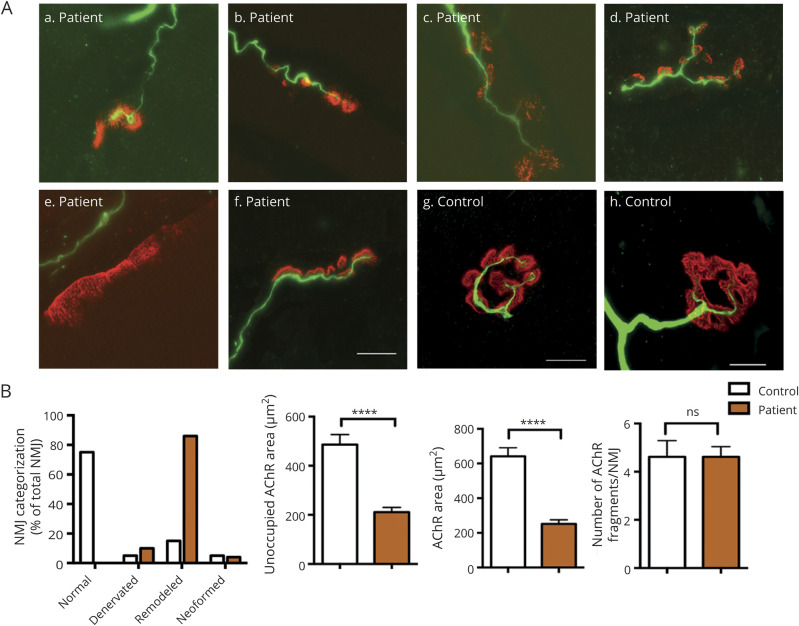
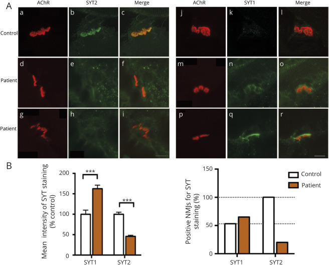
Results: The 2 infants presented a similar clinical phenotype evoking first a congenital myopathy characterized by muscle weakness and hypotonia. Next-generation sequencing allowed to the identification of 1 homozygous intronic mutation c.465+1G>A in patient 1 and another homozygous frameshift mutation c.328_331dup in patient 2, located respectively in the 5' splice donor site of SYT2 intron 4 and in exon 3. Functional studies of the intronic mutation validated the abolition of the splice donor site of exon 4 leading to its skipping. In-frame skipping of exon 4 that encodes part of the C2A calcium-binding domain of SYT2 is associated with a loss-of-function effect resulting in a decrease of neurotransmitter release and severe pre- and postsynaptic NMJ defects.

Conclusions: This study identifies new homozygous recessive SYT2 mutations as the underlying cause of severe and early presynaptic form of CMS expanding the genetic spectrum of recessive SYT2-related CMS associated with defects in neurotransmitter release.


Figures

Figure 1
Figure 1. Genetic and electrophysiologic features of recessive SYT2 mutations
(A) The family 1 and 2 pedigrees revealed that only patients 1 and 2 (in black) are affected in the families and support the recessive autosomal heredity from consanguineous families at second degree with grandparents who are sister and brother in both cases. (B) Decrement at RNS (3 Hz) was observed from 25% to 39% in 3 muscles/nerve. (C) Image of patient 2 showing a severe congenital hypotonia with muscle weakness preventing him from holding his head, respiratory failure requiring NIV, and a constant feeding tube. (D) Position of the identified mutations on the structure of SYT2 protein. Intronic and frameshift mutations identified in this study are indicated in red, respectively, in the C2A domain and in exon 3, whereas the homozygote recessive mutation described in the literature is noted in underlined red. The dominant missense mutations described in the C2B domain of SYT2 are represented in black. Exons encoding C2A and C2B domains are underlined. The codon START is indicated at the beginning of exon 2; exon 1 does not encode for any amino acids. (E) Sanger sequencing revealed a 465+1G>A substitution in patient 1 cDNA compared with the control. Red: the 4 nucleotides of the control (GACA), which are duplicated in patient 2. Green: modified amino acids. (F) Sequence alignment of the SYT2 region corresponding to nucleotides 319 to 354 (amino acids 107–118) of patient 2 compared with the control. Red: the 4 nucleotides of the control (GACA), which are duplicated in patient 2. Green: modified amino acids. *Premature stop codon. NIV = noninvasive ventilation.
Figure 2
Figure 2. Patient 1 exon 4 skipping using the splicing reporter minigene PCAS2
(A) Schematic representation of the pCAS2-SYT2-exon4. Genomic DNA of the patient and the control has been inserted into natural intron 3 of SERPING1/CINH gene surrounded by exons 2 and 3. Arrows: primers used for RT-PCR analysis. (B) After transfection in HEK293T cells and RT-PCR analysis of the minigene transcripts containing control or patient genomic DNA, agarose gel electrophoresis shows that the c.465+1G>A mutation induces the loss of 120 nucleotides corresponding to the complete patient's exon 4 skipping. Resulting PCR products are labeled exon 4 for the control (520 bp) and ∆ exon 4 for the patient (400 bp). GAPDH, amplified to confirm equal amounts of starting cDNA, shows comparable transcripts quantity between the patient and the control.
Figure 3
Figure 3. Morphologic analysis of NMJ in patient 1
(A) Representative NMJ confocal pictures of the patient muscle biopsy immunostained with an anti-neurofilament antibody in green to label the motor axons and with α-bungarotoxin in red for postsynaptic AChR. Representative control, neoformed, remodeled, and denervated NMJ pictures are shown. Scale bar: 10 μm. (B) Histogram of NMJs classification into 4 categories and postsynaptic morphometric analyses. Changes in NMJs distribution with a majority of remodeled NMJs were observed in the patient compared with the control. Using human-adapted NMJ-morph workflow analysis on confocal z-stack projections of individual NMJs, histograms of postsynaptic components such as AChR fragment number, AChR area, or unoccupied AChR area showed a major remodeling of NMJs in the patient compared with the control. The same parameters were applied to the patient and the control. ****p < 0.0001. All error bars are SEM. AChR = acetylcholine receptor; NMJ = neuromuscular junction; ns = no significant change (p > 0.05).
Figure 4
Figure 4. Expression level of SYT-1 and -2 in patient 1
(A) Immunostaining study of SYT1 and SYT2 in green together with AChR in red to visualize the synaptic area. All NMJs express SYT2 in the control (a–c), whereas SYT2 staining intensity is decreased in the patient's NMJs (d–i). A large majority of NMJs expressing a low immunostaining intensity of SYT1 were detected in the control (j–l) while an increase in staining intensity and number of NMJs expressing SYT1 was observed in patient's samples (m–r). Scale bar: 10 μm. (B) Histograms of mean intensity and percentage of positive NMJs for SYT1 and SYT2 staining on confocal z-stack projections of all patient's NMJs analyzed, including negative immunostaining, compared with the control. Number of NMJs analyzed: 50 for the patient and 30 for the control. ***p < 0.0001. All error bars are SEM. AChR = acetylcholine receptor; NMJ = neuromuscular junction; SYT = synaptotagmin.

References

    1. Tintignac LA, Brenner HR, Rüegg MA. Mechanisms regulating neuromuscular junction development and function and causes of muscle wasting. Physiol Rev 2015;95:809–852. - PubMed
    1. Rodríguez Cruz PM, Palace J, Beeson D. The neuromuscular junction and wide heterogeneity of congenital myasthenic syndromes. Int J Mol Sci 2018;19:1677. - PMC - PubMed
    1. Nicole S, Azuma Y, Bauché S, Eymard B, Lochmüller H, Slater C. Congenital myasthenic syndromes or inherited disorders of neuromuscular transmission: recent discoveries and open questions. J Neuromuscul Dis 2017;4:269–284. - PMC - PubMed
    1. Engel AG. Congenital myasthenic syndromes in 2018. Curr Neurol Neurosci Rep 2018;18:46. - PubMed
    1. Lee J, Guan Z, Akbergenova Y, Littleton JT. Genetic analysis of synaptotagmin C2 domain specificity in regulating spontaneous and evoked neurotransmitter release. J Neurosci 2013;33:187–200. - PMC - PubMed