

Changes in connexin 43 in inflammatory skin disorders: Eczema, psoriasis, and Steven-Johnson syndrome/toxic epidermal necrolysis
- PMID: 33659713
- PMCID: PMC7895532
- DOI: 10.1002/hsr2.247
Changes in connexin 43 in inflammatory skin disorders: Eczema, psoriasis, and Steven-Johnson syndrome/toxic epidermal necrolysis
Abstract
Background: Connexin 43 (Cx43) plays a central role in the inflammatory response and wound healing. Targeting Cx43 expression reduces inflammation in a variety of injuries. The expression pattern of Cx43 has not been described for many inflammatory skin diseases.
Objectives: To describe the expression patterns of Cx43 in eczema, psoriasis, Steven-Johnson syndrome/toxic epidermal necrolysis.
Methods: Archival skin biopsies from patients with eczema, psoriasis, and Steven-Johnson syndrome/toxic epidermal necrosis were identified and examined, with sister sections stained for Cx43 and imaged by confocal microscopy. All samples were compared to age and site-matched normal skin controls.
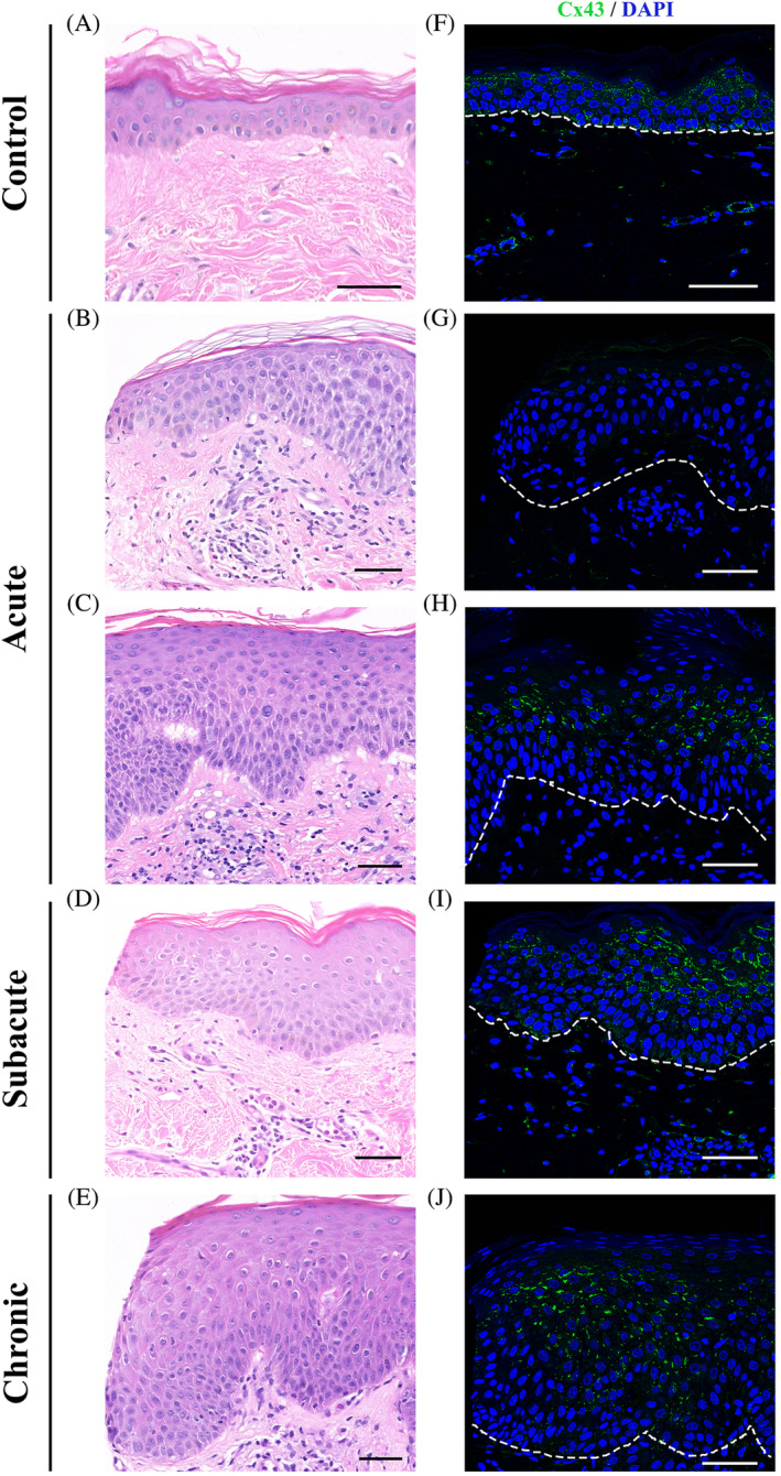
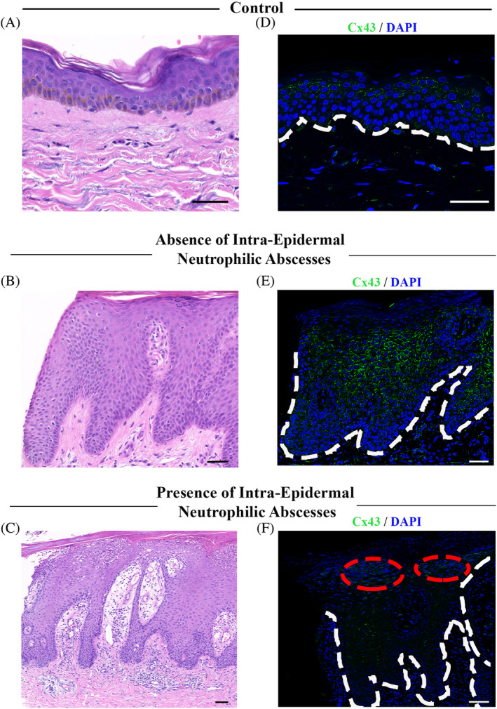
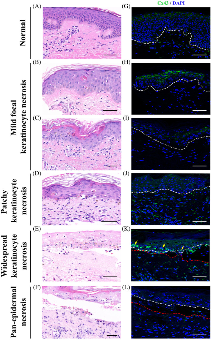
Results: Epidermal Cx43 is reduced in acute eczema, absent in regions of spongiosis, and is highly elevated in subacute and chronic eczema. In plaque psoriasis, Cx43 is overexpressed in areas with psoriasiform hyperplasia with a fish-scale-like appearance but is lost in regions surrounding neutrophil microabscesses. Cx43 staining is strong in the neutrophils within these microabscesses. In SJS/TEN, Cx43 expression is elevated in areas bordering normal tissue but is rapidly lost in areas of keratinocyte necrosis.
Conclusions: Dynamic changes in Cx43 levels are seen in inflammatory skin diseases and may represent future potential therapeutic targets.
Keywords: dermatitis; epidermal necrosis; expression patterns; gap junction; psoriasiform.
© 2021 The Authors. Health Science Reports published by Wiley Periodicals LLC.
Conflict of interest statement
The authors have no conflict of interest to declare.
Figures
References
-
- Alexander DB, Goldberg GS. Transfer of biologically important molecules between cells through gap junction channels. Curr Med Chem. 2003;10:2045‐2058. - PubMed
-
- Churko JM, Laird DW. Gap junction remodeling in skin repair following wounding and disease. Physiology (Bethesda). 2013;28:190‐198. - PubMed
-
- Salomon D, Masgrau E, Vischer S. Topography of mammalian connexins in human skin. J Invest Dermatol. 1994;103:240‐247. - PubMed
-
- Van Steensel MA, van Geel M, Nahuys MA, et al. Novel connexin 26 mutation in a patient diagnosed with keratitis‐ichthyosis‐deafness syndrome. J Invest Dermatol. 2002. Apr;118:724‐727. - PubMed
-
- Huang T, Shao Q, MacDonald A, et al. Autosomal recessive GJA1 (Cx43) gene mutations cause oculodentodigital dysplasia by distinct mechanisms. J Cell Sci. 2013. Jul 1;126:2857‐2866. - PubMed
LinkOut - more resources
Full Text Sources
Other Literature Sources
Miscellaneous
