Review
. 2021 Jan 19:10:4.
doi: 10.12703/r/10-4. eCollection 2021.

The cancer cell secretome drives cooperative manipulation of the tumour microenvironment to accelerate tumourigenesis

Affiliations
Review

The cancer cell secretome drives cooperative manipulation of the tumour microenvironment to accelerate tumourigenesis

Shona Ritchie et al. Fac Rev. .

Abstract

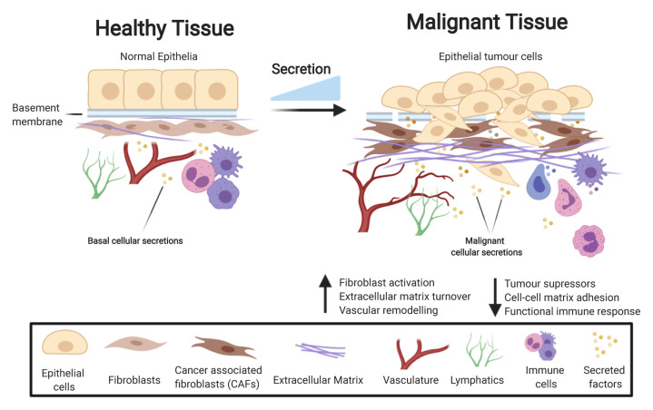
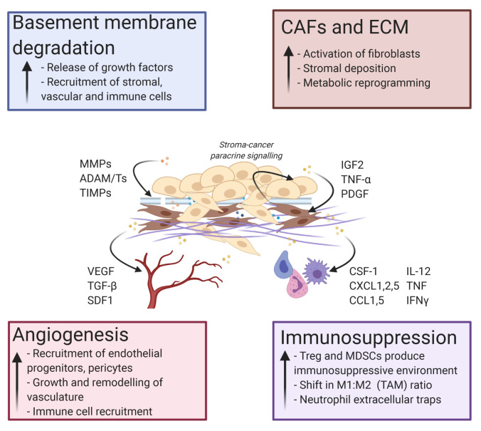
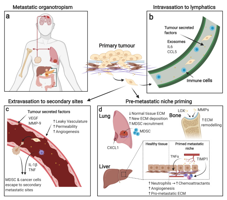
Cellular secretions are a fundamental aspect of cell-cell and cell-matrix interactions in vivo. In malignancy, cancer cells have an aberrant secretome compared to their non-malignant counterparts, termed the "cancer cell secretome". The cancer cell secretome can influence every stage of the tumourigenic cascade. At the primary site, cancer cells can secrete a multitude of factors that facilitate invasion into surrounding tissue, allowing interaction with the local tumour microenvironment (TME), driving tumour development and progression. In more advanced disease, the cancer cell secretome can be involved in extravasation and metastasis, including metastatic organotropism, pre-metastatic niche (PMN) preparation, and metastatic outgrowth. In this review, we will explore the latest advances in the field of cancer cell secretions, including its dynamic and complex role in activating the TME and potentiating invasion and metastasis, with comments on how these secretions may also promote therapy resistance.

Keywords: Cancer cell secretome; Pre-metastatic niche; Stroma; Tumour microenvironment.


Conflict of interest statement

The authors declare that they have no competing interests.No competing interests were disclosed.No competing interests were disclosed.Anil K. Sood consults for Merck, Astra Zeneca, and Kiyatec, he is a shareholder for BioPath, and receives research funding from M Trap.

Figures

Figure 1.
Figure 1.. The cancer cell secretome drives a pro-tumourigenic environment.
During the process of cancer development, the secretome is markedly changed compared to healthy tissue, with increased levels of secretion resulting in a change to many key processes enhancing tumour growth. Examples of pathways affected by tumour cell-derived secretion include an increase in fibroblast activation, extracellular matrix (ECM) deposition, and vascular remodelling while key pathways in tumour suppression, cell–cell matrix adhesion, and regulation of the immune system are lost.
Figure 2.
Figure 2.. The cancer cell secretome drives tumour microenvironmental changes enhancing tumour growth, invasion, and metastasis.
Tumour cell secretion activates a number of pro-tumourigenic processes. These events include basement membrane degradation, activation of cancer-associated fibroblasts (CAFs), deposition of extracellular matrix (ECM), angiogenesis at the tumour site, and immunomodulation to favour an immunosuppressive environment. Examples of key tumour-secreted proteins that affect each major process are shown. ADAM/T, a disintegrin and metalloproteinase/with thrombospondin motif; CCL, C-C motif chemokine ligand; CSF-1, colony-stimulating factor 1; CXCL, C-X-C motif chemokine ligand; IFN, interferon; IGF2, insulin-like growth factor 2; IL, interleukin; MDSC, myeloid-derived suppressor cell; MMP, matrix metalloproteinase; PDGF, platelet-derived growth factor; SDF1, stromal cell-derived factor 1; TAM, tumour-associated macrophage; TGF, transforming growth factor; TIMP, tissue inhibitor of metalloproteinase; TNF, tumour necrosis factor; Treg, regulatory T cell; VEGF, vascular endothelial growth factor.
Figure 3.
Figure 3.. The tumour secretome activates pro-metastatic events.
Successful tumour cell metastasis and survival at secondary sites is a multi-step process involving many different mechanisms. (a) Tumour cells originating from different primary tumour sites display organotropism for secondary metastasis sites; examples of this include breast cancer’s favourable growth in the brain, bone, and lung as well as pancreatic and colon cancer’s favourable growth in the liver. (b) Tumour cells initially preferentially metastasise to the lymphatic system owing to a favourable environment where they can then spread to distant secondary organs. (c) Tumour-derived secreted proteins can provide pro-metastatic signals during vasculature remodelling. For example, tumour-secreted proteins can induce leaky vasculature, increasing permeability for tumour cells and other cells to access secondary sites. (d) Metastatic niche priming can also be affected by primary tumour secretions, with examples of priming including extracellular matrix (ECM) remodelling, immune system recruitment, vascular remodelling, and chemoattractant secretion at metastatic sites. CCL5, C-C motif chemokine ligand 5; CXCL1, C-X-C motif chemokine ligand 1; IL, interleukin; LOX, lysyl oxidase; MDSC, myeloid-derived suppressor cell; MMP, matrix metalloproteinase; TIMP, tissue inhibitor of metalloproteinase; TNF, tumour necrosis factor; VEGF, vascular endothelial growth factor.

References

    1. Tjalsma H, Bolhuis A, Jongbloed JD, et al. : Signal peptide-dependent protein transport in Bacillus subtilis: A genome-based survey of the secretome. Microbiol Mol Biol Rev. 2000; 64(3): 515–47. 10.1128/mmbr.64.3.515-547.2000 - DOI - PMC - PubMed
    1. Zhang M, Schekman R: Cell biology. Unconventional secretion, unconventional solutions. Science. 2013; 340(6132): 559–61. 10.1126/science.1234740 - DOI - PubMed
    1. Kim J, Gee HY, Lee MG: Unconventional protein secretion - new insights into the pathogenesis and therapeutic targets of human diseases. J Cell Sci. 2018; 131(12): jcs213686. 10.1242/jcs.213686 - DOI - PubMed
    1. Uhlén M, Karlsson MJ, Hober A, et al. : The human secretome. Sci Signal. 2019; 12(609): eaaz0274. 10.1126/scisignal.aaz0274 - DOI - PubMed
    2. Faculty Opinions Recommendation

    1. Zhang H, Freitas D, Kim HS, et al. : Identification of distinct nanoparticles and subsets of extracellular vesicles by asymmetric flow field-flow fractionation. Nat Cell Biol. 2018; 20(3): 332–43. 10.1038/s41556-018-0040-4 - DOI - PMC - PubMed
    2. Faculty Opinions Recommendation

LinkOut - more resources