Review
. 2020 Nov 25:9:17.
doi: 10.12703/r/9-17. eCollection 2020.

New techniques for studying neurodevelopment

Affiliations
Review

New techniques for studying neurodevelopment

Augusto Escalante et al. Fac Rev. .

Abstract

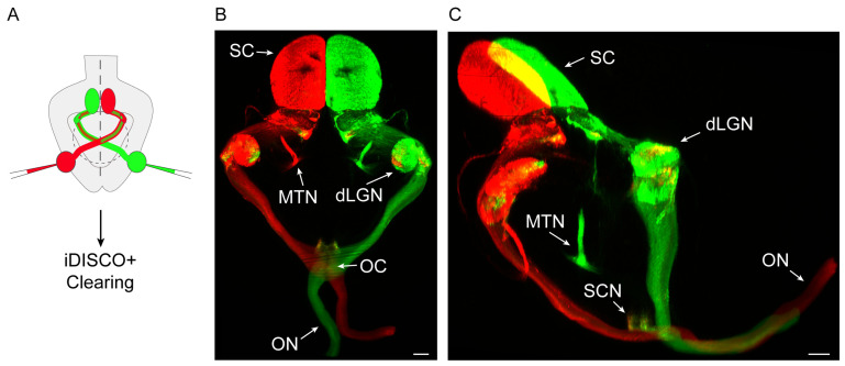
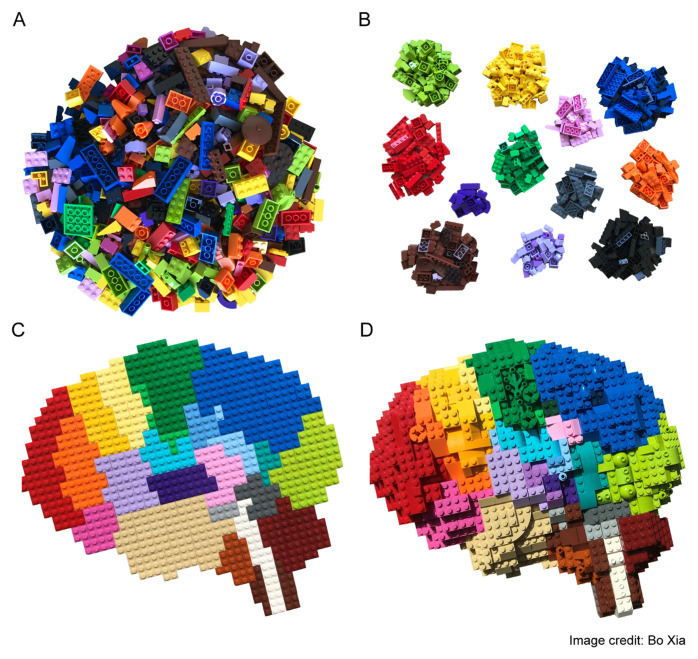
The extraordinary diversity, variability, and complexity of cell types in the vertebrate brain is overwhelming and far exceeds that of any other organ. This complexity is the result of multiple cell divisions and intricate gene regulation and cell movements that take place during embryonic development. Understanding the cellular and molecular mechanisms underlying these complicated developmental processes requires the ability to obtain a complete registry of interconnected events often taking place far apart from each other. To assist with this challenging task, developmental neuroscientists take advantage of a broad set of methods and technologies, often adopted from other fields of research. Here, we review some of the methods developed in recent years whose use has rapidly spread for application in the field of developmental neuroscience. We also provide several considerations regarding the promise that these techniques hold for the near future and share some ideas on how existing methods from other research fields could help with the analysis of how neural circuits emerge.

Keywords: Clearing; Light sheet microscopy; Machine learning; Neural development tools; scRNAseq.


Conflict of interest statement

The authors declare that they have no competing interests.No competing interests were disclosed.No competing interests were disclosed.

Figures

Figure 1.
Figure 1.. Three-dimensional (3D) view of retinal axons projecting to the visual nuclei within the mouse brain.
A. Scheme of the experimental approach. A postnatal mouse is injected with fluorescent tracers of different colours in each eye and then processed through the iDISCO+ clearing protocol. B. Dorsal view of a light sheet fluorescence microscope (LSFM)-acquired 3D image stack from the whole brain of a mouse injected with different colour tracers into each eye. dLGN, dorsal lateral geniculate nucleus; OC, optic chiasm; ON, optic nerve; MTN, medial terminal nucleus; SC, superior colliculus. Scale bar: 300 µm. C. Mediolateral view of an LSFM-acquired 3D image stack from the whole brain of a mouse injected with different colour tracers into each eye. dLGN, dorsal lateral geniculate nucleus; MTN, medial terminal nucleus; ON, optic nerve; SC, superior colliculus; SCN, suprachiasmatic nucleus. Scale bar: 300 µm.
Figure 2.
Figure 2.. Building-block representation of single cell transcriptome modalities.
A. Bulk RNA sequencing (RNAseq) experiments use a large number of cells as starting material, which results in a high depth and resolution at the transcriptomic level. However, because the measurements obtained represent an average of gene expression across all of the cells present in the sample, any differences between cells become occluded. B. Single cell RNAseq (scRNAseq) methods are capable of maintaining cell individuality during isolation of mRNA molecules. mRNAs are tagged and reconstructed informatically so that they can be assigned to a particular cell. This enables the identification of cell clusters according to their transcriptomic signatures, but spatial information is still lost. C. The spatial location of each cell is maintained in spatial transcriptomics approaches. By fluorescently tagging each mRNA species or recording the position in situ with barcodes, spatial information may be assigned to each cell together with their transcriptomic profile. Currently, the best attainable resolution is within the tens of microns range, which is still far from ideal and sensitivity remains low. As this is a novel method, the availability of the protocol is scarce and its adoption outside originator labs is therefore difficult. D. Researchers are now advancing towards an integrated (genetic, transcriptomic, and proteomic) representation of the brain in time and space. This figure has been reused with permission from the creator Bo Xia.

References

    1. The Cajal Legacy: Consejo Superior de Investigaciones Científicas - CSIC - csic.es Reference Source
    1. Alain Chédotal en Twitter: ‘It was a risky experiment but thanks to Ripley we did it: this is our contribution to #AlienDay enjoy https://t.co/QyZHFtfkYy’ / Twitter. Twitter https://twitter.com/alainchedotal/status/1254456825514254337.
    1. Dent JA, Polson AG, Klymkowsky MW: A whole-mount immunocytochemical analysis of the expression of the intermediate filament protein vimentin in Xenopus. Development. 1989; 105(1): 61–74. - PubMed
    1. Keller PJ, Dodt HU: Light sheet microscopy of living or cleared specimens. Curr Opin Neurobiol. 2012; 22(1): 138–43. 10.1016/j.conb.2011.08.003 - DOI - PubMed
    1. Voie AH, Burns DH, Spelman FA: Orthogonal-plane fluorescence optical sectioning: Three-dimensional imaging of macroscopic biological specimens. J Microsc. 1993; 170(Pt 3): 229–36. 10.1111/j.1365-2818.1993.tb03346.x - DOI - PubMed