. 2021 Mar;473(3):557-571.
doi: 10.1007/s00424-021-02537-y. Epub 2021 Mar 4.

The conduction velocity-potassium relationship in the heart is modulated by sodium and calcium

Affiliations

The conduction velocity-potassium relationship in the heart is modulated by sodium and calcium

D Ryan King et al. Pflugers Arch. 2021 Mar.

Abstract

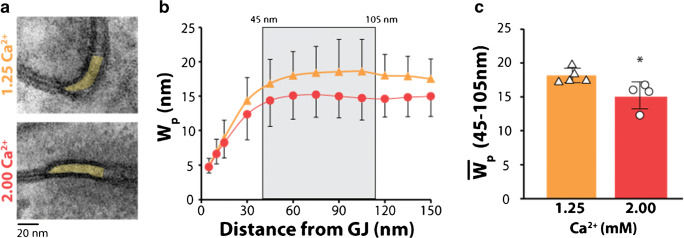
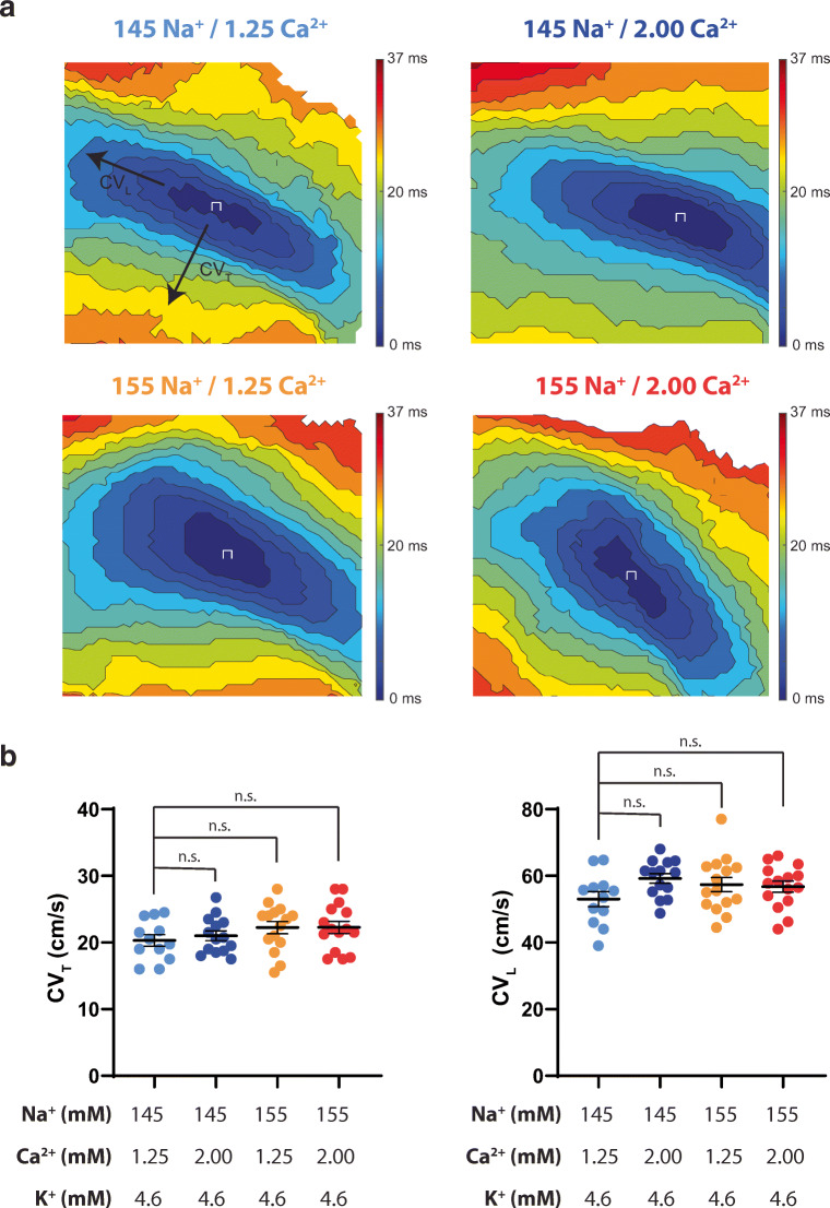
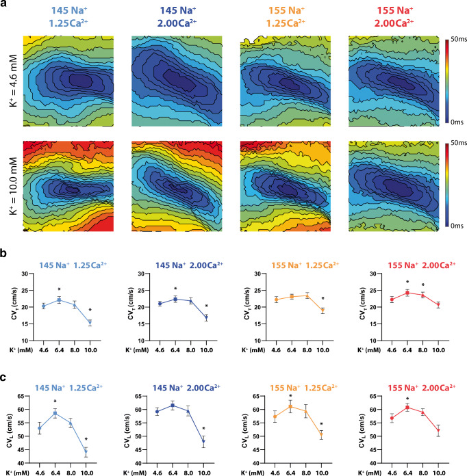
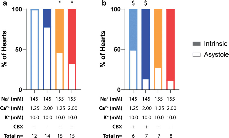
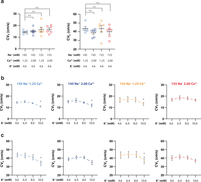
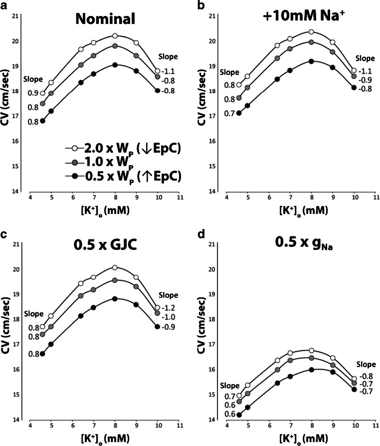
The relationship between cardiac conduction velocity (CV) and extracellular potassium (K+) is biphasic, with modest hyperkalemia increasing CV and severe hyperkalemia slowing CV. Recent studies from our group suggest that elevating extracellular sodium (Na+) and calcium (Ca2+) can enhance CV by an extracellular pathway parallel to gap junctional coupling (GJC) called ephaptic coupling that can occur in the gap junction adjacent perinexus. However, it remains unknown whether these same interventions modulate CV as a function of K+. We hypothesize that Na+, Ca2+, and GJC can attenuate conduction slowing consequent to severe hyperkalemia. Elevating Ca2+ from 1.25 to 2.00 mM significantly narrowed perinexal width measured by transmission electron microscopy. Optically mapped, Langendorff-perfused guinea pig hearts perfused with increasing K+ revealed the expected biphasic CV-K+ relationship during perfusion with different Na+ and Ca2+ concentrations. Neither elevating Na+ nor Ca2+ alone consistently modulated the positive slope of CV-K+ or conduction slowing at 10-mM K+; however, combined Na+ and Ca2+ elevation significantly mitigated conduction slowing at 10-mM K+. Pharmacologic GJC inhibition with 30-μM carbenoxolone slowed CV without changing the shape of CV-K+ curves. A computational model of CV predicted that elevating Na+ and narrowing clefts between myocytes, as occur with perinexal narrowing, reduces the positive and negative slopes of the CV-K+ relationship but do not support a primary role of GJC or sodium channel conductance. These data demonstrate that combinatorial effects of Na+ and Ca2+ differentially modulate conduction during hyperkalemia, and enhancing determinants of ephaptic coupling may attenuate conduction changes in a variety of physiologic conditions.

Keywords: Calcium; Cardiac Electrophysiology; Hyperkalemia; Langendorff; Sodium.


Figures

Fig. 1
Fig. 1
Increasing Ca2+ decreases Wp in the presence of 155-mM Na+. (a) Representative transmission electron micrographs of perinexi with 1.25- and 2.00-mM Ca2+, (b) Wp measurements from 0 to 150 nm from GJ plaque, (c) Average Wp measurements for 45–105 nm from GJ plaque, Wp significantly decreased with 2.00-mM Ca2+. (n = 5 hearts perfused with 1.25-mM Ca+2 and n = 4 hearts with 2.00-mM Ca2+, *p < 0.05 compared to 1.25-mM Ca2+ via two-tailed Student’s t test)
Fig. 2
Fig. 2
Altering Na+, Ca2+, or Na+ and Ca2+ does not change CVT or CVL at 4.6-mM K+. (a) Representative isochrone maps for each Na+/Ca2+ perfusion combination. The 145-mM Na+/1.25-mM Ca2+ map is marked with CVT and CVL designations for visualization purposes, (b) Summary of CVT and CVL at 4.6-mM K+. p < 0.05 denoted by *, significance determined by one-way ANOVA with Dunnett’s correction for multiple comparisons. (n = 12, 14, 15, 15 from left to right respectively for both CVT and CVL)
Fig. 3.
Fig. 3.
Simultaneously increasing Na+ and Ca2+ preserves CVT and CVL at 10.0-mM K+. (a) Representative isochrone maps for each Na+/Ca2+ perfusion combination at 4.6- and 10.0-mM K+, (b) Summary of CVT as a function of K+ for all Na+ and Ca2+ perfusion combinations, (c) Summary of CVL as a function of K+ for all Na+ and Ca2+ perfusion combinations. p < 0.05 denoted by *, significance determined by one-way ANOVA with Dunnett’s correction for multiple comparisons (n = 12, 14, 15, 15 from left to right, respectively).
Fig. 4
Fig. 4
Incidence of asystole during 10.0-mM K+ perfusion. (a) Elevating perfusate Na+ significantly increases the preservation of intrinsic rhythm at 10.0-mM K+ perfusion (significance determined by Fisher’s exact test; * denotes p < 0.05 as compared to 145-mM Na+, 1.25-mM Ca2+, 10.0-mM K+ perfusate group), (b) Inhibiting GJs with CBX (30 μM) significantly increases the preservation of intrinsic rhythm in the presence of 145-mM Na+ when compared to the control condition (significance determined by Fisher’s exact test; $ denotes p < 0.05 compared to each perfusates’ respective CBX – group). There were no significant differences in preservation of intrinsic rhythm across groups perfused with CBX
Fig. 5
Fig. 5
Following GJ inhibition with CBX, simultaneously increasing Na+ and Ca2+ preserves CVT and CVL at 10.0-mM K+. (a) Altering Na+, Ca2+, or Na+ and Ca2+ does not change CVT or CVL at 4.6-mM K+, (b) Summary of CVT as a function of K+ in the presence of CBX (n = 6, 7, 7, 8 from left to right, respectively), (c) Summary of CVL as a function of K+ in the presence of CBX (n = 6, 7, 7, 8 from left to right, respectively). Significance determined by ordinary one-way ANOVA with Dunnett’s correction for multiple comparisons (p < 0.05 denoted by *)
Fig. 6
Fig. 6
Computational predictions of modulating perinexal width (WP), extracellular sodium concentrations (Na+), gap junctional coupling (GJC), and the fast sodium channel conductance (gNa). (a) Increasing WP to reduce EpC is associated with increased conduction velocity (CV, black to white points). The positive slope calculated from a linear fit of CV over the range of extracellular potassium (K+) from 4.56 to 7 mM is reduced as WP decreases. The negative slope associated with sodium channel inactivation over the range of 9- to 10-mM K+ decreases to a greater extent with narrow WP, (b) Increasing Na+ by 10 mM (+10-mM Na+) decreases the positive slope of the CV-K+ relationship without substantively altering the negative slope or CV overall, (c) Reducing GJC by 50% (0.5 × GJC) slows CV for all values of K+ without changing the positive CV-K+ slope. However, 0.5 × GJC is associated with enhanced CV slowing during sodium channel inactivation measured between 9- and 10-mM K+ relative to the nominal condition in panel b. CV slowing was still the lowest with the narrowest cleft widths (0.5 × WP), (d) Reducing gNa by 50% (0.5 × gNa) not only reduces CV for all K+, but it also reduces both the positive and negative slopes of the CV-K+ relationship, without altering predictions that WP associated with the slowest CV attenuates CV-dependent changes on K+

References

    1. Aiba T, Hesketh GG, Liu T, Carlisle R, Villa-Abrille MC, O'Rourke B, Akar FG, Tomaselli GF. Na+ channel regulation by Ca2+/calmodulin and Ca2+/calmodulin-dependent protein kinase II in guinea-pig ventricular myocytes. Cardiovasc Res. 2010;85(3):454–463. doi: 10.1093/cvr/cvp324. - DOI - PMC - PubMed
    1. Antzelevitch C. Brugada syndrome. PACE - Pacing Clin Electrophysiol. 2006;29(10):1130–1159. doi: 10.1111/j.1540-8159.2006.00507.x. - DOI - PMC - PubMed
    1. Ashpole NM, Herren AW, Ginsburg KS, Brogan JD, Johnson DE, Cummins TR, Bers DM, Hudmon A. Ca 2+/calmodulin-dependent protein kinase II (CaMKII) regulates cardiac sodium channel Na V 1.5 gating by multiple phosphorylation sites. J Biol Chem. 2012;287(24):19856–19869. doi: 10.1074/jbc.M111.322537. - DOI - PMC - PubMed
    1. Bartos DC, Grandi E, Ripplinger CM. Ion channels in the heart. Compr Physiol. 2015;5(3):1423–1464. doi: 10.1002/cphy.c140069. - DOI - PMC - PubMed
    1. Bates D, Mächler M, Bolker B, Walker S (2015) Fitting linear mixed-effects models using lme4. J Stat Softw 67(1). 10.18637/jss.v067.i01

Publication types