Case Reports
. 2022 Jun;25(2):369-377.
doi: 10.1007/s40477-021-00572-0. Epub 2021 Mar 3.

Isolated partial tear of extensor digitorum longus tendon with overlying muscle herniation in acute ankle sports injury: role of high resolution musculoskeletal ultrasound

Affiliations
Case Reports

Isolated partial tear of extensor digitorum longus tendon with overlying muscle herniation in acute ankle sports injury: role of high resolution musculoskeletal ultrasound

Jeena Bordoloi Deka et al. J Ultrasound. 2022 Jun.

Abstract

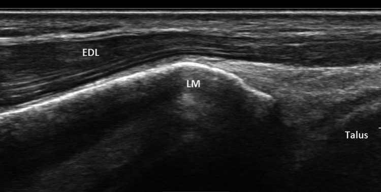
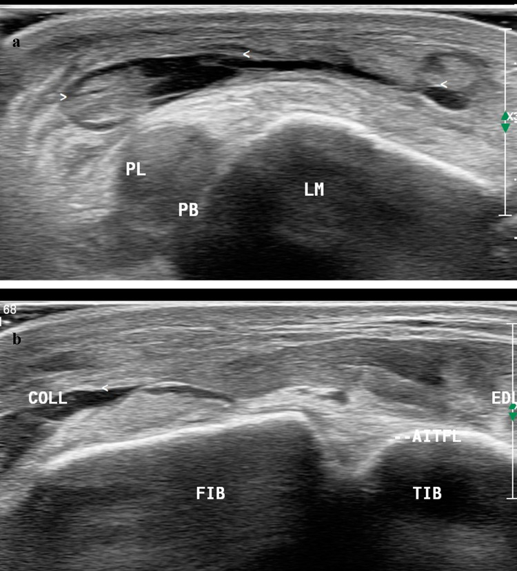
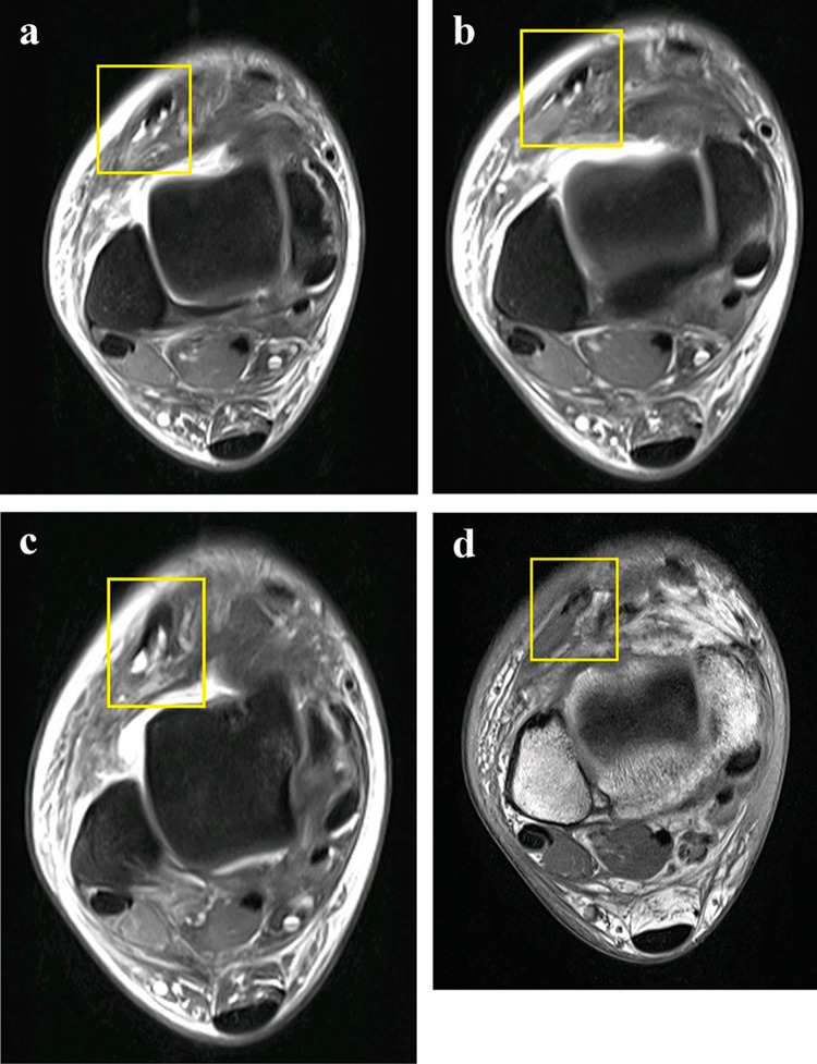
Lateral Ankle sprain is a common sports-related trauma with the mechanism of injury ranging from inversion to plantar flexion. These injuries commonly affect the ligaments but can also affect the associated soft tissue structures like the eversion muscles and tendons. Prompt and accurate diagnosis of such injuries is warranted so as to ensure early return to play and prevent long-term complications. Lateral ankle sprain injuries in sports may not always be associated with ligament injuries. We report a never before reported case of lateral ankle sprain injury in a soccer player with the unusual finding of isolated partial tear of Extensor digitorum longus muscle and its fascia leading to myo-fascial herniation. The lateral ankle ligaments were intact. The diagnosis was clinched on a high-frequency ultrasound scan supported by dynamic maneuvers which in fact proved to be superior to MRI as the latter failed to demonstrate the myo-fascial herniation in our case. We therefore propose that real-time ultrasound scanning with dynamic maneuvers should be the first line of investigation to assess sports injuries in anatomically complex joints like the ankle.

Keywords: Ankle sports injury; Extensor digitorum longus tear; High-resolution musculoskeletal ultrasound; Inversion and plantar flexion injuries; Lateral ankle sprain; Muscle hernia.


Conflict of interest statement

CB is a consultant for Bracco Imaging and Doc. Congress. The other authors has nothing to disclose.

Figures

Fig. 1
Fig. 1
Antero-posterior and lateral Radiograph of the ankle joint. Osseous anatomy is normal
Fig. 2
Fig. 2
Long axis of the Extensor Digitorum longus (EDL) at rest. Disruption of the echogenic fascia (arrow) at the level of the lateral malleolus (LM). Herniation of muscle tissue is noted into subcutaneous soft tissue
Fig. 3
Fig. 3
Short axis scan shows the gap in the fascia (epimysium) of the Extensor Digitorum Longus (EDL) shown by dotted line
Fig. 4
Fig. 4
Long axis scan of Extensor digitorum longus (EDL) during dynamic study ie Dorsiflexion. Decrease in size of hernia through defect is noted (arrow)
Fig. 5
Fig. 5
Long axis scan of the Extensor digitorum longus (EDL) over the lateral malleolus (LM). Partial tears of few fibers of the EDL (arrows) superior and inferior to facial tear and hernia level and near MTJ. Small hematomas are noted at the site of tears
Fig. 6
Fig. 6
Long axis scan of the Extensor digitorum longus (EDL) shows bulge and waviness (arrow) in deeper aspect distal to the tears (arrowheads) and site of hernia
Fig. 7
Fig. 7
Long axis scan of normal contralateral Extensor digitorum longus (EDL) over the lateral malleolus (LM) and anterior talar recess shows continuity of the muscle and tendon fibers and intact echogenic fascia (epimysium)
Fig. 8
Fig. 8
Long axis scan over the level of lateral malleolus (LM) shows a subtle fracture (FR) with step-off deformity
Fig. 9
Fig. 9
a, b Short axis scan of the anterior and lateral ankle at rest shows a well-defined collection suggestive of hematoma (arrow head) in the subcutaneous overlying the peroneal longus and brevis tendons (PL, PB). Subcutaneous edema is noted in the anterior and lateral aspect of the ankle. (AITFL: anterior inferior tibiofibular ligament; Tib: Tibia; Fib: Fibula; Coll: collection /hematoma)
Fig. 10
Fig. 10
MRI T1W (ac) and PDFS (d) axial images showing heterogenicity of the Extensor Digitorum longus (yellow box) and partial tear near the Myo-tendinous junction. The epimysium is inconspicuous. The hernia is not seen

References

    1. Pointinger H, et al. Rupture of the extensor digitorum longus muscle: additional finding in an ankle Sprain. European J Trauma. 2003;29:161–163. doi: 10.1007/s00068-003-1270-z. - DOI
    1. Guermazi A, et al. Imaging of muscle injuries in sports medicine: sports imaging series. Radiology. 2017;285:3. doi: 10.1148/radiol.2017174038. - DOI - PubMed
    1. Hattori K, et al. Closed rupture of the extensor digitorum longus tendon—a case report and biochemical analysis of rupture mechanism. The Foot. 2007;17:220–223. doi: 10.1016/j.foot.2007.04.003. - DOI
    1. Konradsen L, et al. Ankle inversion injuries. The role of the dynamic defense mechanism. Am J Sports Med. 1997;25:54–58. doi: 10.1177/036354659702500110. - DOI - PubMed
    1. Harrington AC, Mellette JR., Jr Hernias of the anterior tibialis muscle: case report and review of the literature. J Am Acad Dermatol. 1990;22:123. doi: 10.1016/S0190-9622(08)80009-6. - DOI - PubMed

Publication types

LinkOut - more resources