

Patterns of local residual disease and local failure after intensity modulated/image guided radiation therapy for sinonasal tumors in dogs
- PMID: 33660342
- PMCID: PMC7995431
- DOI: 10.1111/jvim.16076
Patterns of local residual disease and local failure after intensity modulated/image guided radiation therapy for sinonasal tumors in dogs
Abstract
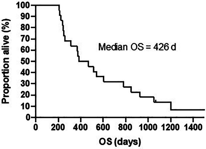
Background: Most dogs with sinonasal tumors (SNT) treated with radiation therapy (RT) died because of local disease progression.
Hypothesis/objectives: Our hypothesis is that the majority of local failure and residual disease would occur within the radiation field.
Animals: Twenty-two dogs with SNT treated with RT.
Methods: Retrospective cohort study.
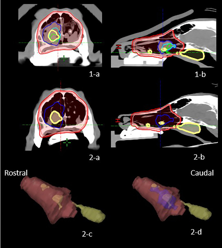
Inclusion criteria: dogs with SNT receiving 10 daily fractions of 4.2 Gy with intensity modulated radiation therapy (IMRT)/image guided radiation therapy (IGRT) and follow-up cone beam computed tomography (CBCT). Each CBCT was registered with the original radiation planning CT and the gross tumor volume (GTV) contoured. The GTV was classified as residual (GTVr) or a failure (GTVf). The dose statistic for each GTV was calculated with the original IMRT plan. For GTVf, failures were classified as "in-field," "marginal," or "out-field" if at least 95, 20-95, or less than 20% of the volume of failure was within 95% (D95) of the total prescription dose, respectively.
Results: There were 52 follow-up CBCT/CTs. Overall there was a GTVr for 20 dogs and GTVf for 16 dogs. The majority of GTVr volume was within the original GTV. GTVf analysis showed that 75% (12/16) were "in-field," 19% (3/16) were "marginal" and 6% (1/16) were "out-field."
Conclusion and clinical importance: In-field failures are the main pattern for local recurrence, and there is evidence of radioresistant subvolumes within the GTV.
Keywords: IGRT; IMRT; dosimetry; nasal tumor.
© 2021 The Authors. Journal of Veterinary Internal Medicine published by Wiley Periodicals LLC on behalf of American College of Veterinary Internal Medicine.
Conflict of interest statement
Authors declare no conflict of interest.
Figures


Similar articles
-
A prospective pilot study on early toxicity from a simultaneously integrated boost technique for canine sinonasal tumours using image-guided intensity-modulated radiation therapy.Vet Comp Oncol. 2018 Dec;16(4):441-449. doi: 10.1111/vco.12399. Epub 2018 May 14. Vet Comp Oncol. 2018. PMID: 29761663
-
Clinical-dosimetric relationship between lacrimal gland dose and keratoconjunctivitis sicca in dogs with sinonasal tumors treated with radiation therapy.J Vet Intern Med. 2020 Mar;34(2):867-872. doi: 10.1111/jvim.15744. Epub 2020 Feb 22. J Vet Intern Med. 2020. PMID: 32086981 Free PMC article.
-
Partial Laryngeal IMRT for T2N0 Glottic Cancer: Impact of Image Guidance and Radiation Therapy Intensification.Int J Radiat Oncol Biol Phys. 2018 Nov 15;102(4):941-949. doi: 10.1016/j.ijrobp.2018.03.034. Epub 2018 Mar 30. Int J Radiat Oncol Biol Phys. 2018. PMID: 29730062
-
INVITED REVIEW--IMAGE REGISTRATION IN VETERINARY RADIATION ONCOLOGY: INDICATIONS, IMPLICATIONS, AND FUTURE ADVANCES.Vet Radiol Ultrasound. 2016 Mar-Apr;57(2):113-23. doi: 10.1111/vru.12342. Epub 2016 Jan 18. Vet Radiol Ultrasound. 2016. PMID: 26777133 Review.
-
Exposure Risks Among Children Undergoing Radiation Therapy: Considerations in the Era of Image Guided Radiation Therapy.Int J Radiat Oncol Biol Phys. 2016 Apr 1;94(5):978-92. doi: 10.1016/j.ijrobp.2015.12.372. Epub 2016 Jan 5. Int J Radiat Oncol Biol Phys. 2016. PMID: 27026304 Review.
Cited by
-
Relative tumor volume has prognostic relevance in canine sinonasal tumors treated with radiation therapy: A retrospective study.PLoS One. 2022 May 27;17(5):e0269083. doi: 10.1371/journal.pone.0269083. eCollection 2022. PLoS One. 2022. PMID: 35622873 Free PMC article.
-
Automatic gross tumor segmentation of canine head and neck cancer using deep learning and cross-species transfer learning.Front Vet Sci. 2023 Mar 21;10:1143986. doi: 10.3389/fvets.2023.1143986. eCollection 2023. Front Vet Sci. 2023. PMID: 37026102 Free PMC article.
-
Evaluation of acute ocular toxicity after definitive-intent radiation therapy in canine sinonasal tumors.PLoS One. 2025 Aug 11;20(8):e0329073. doi: 10.1371/journal.pone.0329073. eCollection 2025. PLoS One. 2025. PMID: 40788897 Free PMC article.
-
Employing deep convolutional neural networks for segmenting the medial retropharyngeal lymph nodes in CT studies of dogs.Vet Radiol Ultrasound. 2022 Nov;63(6):763-770. doi: 10.1111/vru.13132. Epub 2022 Jul 25. Vet Radiol Ultrasound. 2022. PMID: 35877815 Free PMC article.
References
-
- Kent MS, Turek MM, Farrelly J. Recent advances in veterinary radiation oncology. Vet Comp Oncol. 2018;16(2):167‐169. - PubMed
-
- Nolan MW, Gieger TL. Update in veterinary radiation oncology: focus on stereotactic radiation therapy. Vet Clin North Am Small Anim Pract. 2019;49(5):933‐947. - PubMed
-
- Larue SM, Gordon IK. Radiation oncology. In: Vail DM, Thamm D, Liptak J, eds. Withrow and MacEwen's Small Animal Clinical Oncology. St. Louis, Missouri: Elsevier Health Sciences; 2019:215‐218.
-
- Mortier JR, Blackwood L. Treatment of nasal tumours in dogs: a review. J Small Anim Pract. 2020;61(7):404‐415. - PubMed
MeSH terms
Grants and funding
LinkOut - more resources
Full Text Sources
Other Literature Sources
Medical
Research Materials
