. 2021 Apr;20(4):e13331.
doi: 10.1111/acel.13331. Epub 2021 Mar 4.

A novel p53 regulator, C16ORF72/TAPR1, buffers against telomerase inhibition

Affiliations

A novel p53 regulator, C16ORF72/TAPR1, buffers against telomerase inhibition

Yahya Benslimane et al. Aging Cell. 2021 Apr.

Abstract

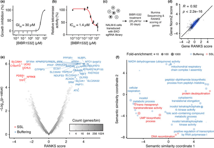
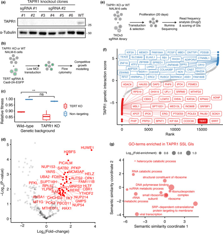
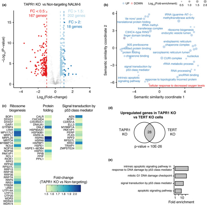
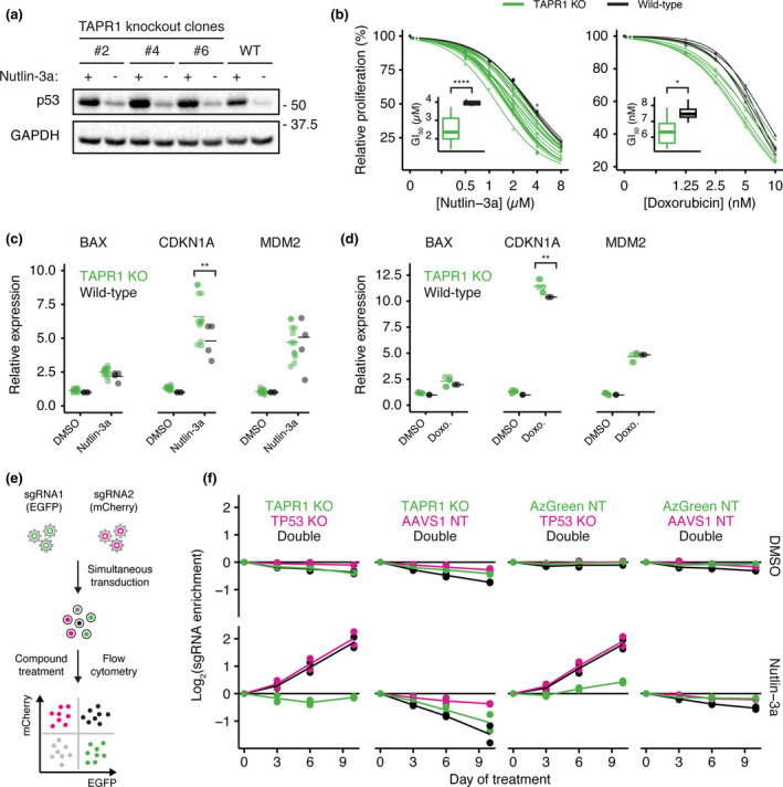
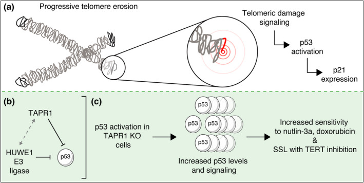
Telomere erosion in cells with insufficient levels of the telomerase reverse transcriptase (TERT), contributes to age-associated tissue dysfunction and senescence, and p53 plays a crucial role in this response. We undertook a genome-wide CRISPR screen to identify gene deletions that sensitized p53-positive human cells to telomerase inhibition. We uncovered a previously unannotated gene, C16ORF72, which we term Telomere Attrition and p53 Response 1 (TAPR1), that exhibited a synthetic-sick relationship with TERT loss. A subsequent genome-wide CRISPR screen in TAPR1-disrupted cells reciprocally identified TERT as a sensitizing gene deletion. Cells lacking TAPR1 or TERT possessed elevated p53 levels and transcriptional signatures consistent with p53 upregulation. The elevated p53 response in TERT- or TAPR1-deficient cells was exacerbated by treatment with the MDM2 inhibitor and p53 stabilizer nutlin-3a and coincided with a further reduction in cell fitness. Importantly, the sensitivity to treatment with nutlin-3a in TERT- or TAPR1-deficient cells was rescued by loss of p53. These data suggest that TAPR1 buffers against the deleterious consequences of telomere erosion or DNA damage by constraining p53. These findings identify C16ORF72/TAPR1 as new regulator at the nexus of telomere integrity and p53 regulation.

Keywords: C16ORF72; CRISPR-Cas9; Telomere Attrition and P53 Response 1; genome-wide screen; p53; synthetic-sick-lethal; telomerase inhibitor (BIBR1532); telomere erosion.


Conflict of interest statement

The authors declare no competing interests.

Figures

FIGURE 1
FIGURE 1
Genome‐wide CRISPR knockout screen identifies chemical‐genetic interactions with telomerase inhibition by BIBR1532. (a) Growth inhibition of NALM‐6 cells upon treatment with the indicated concentrations of BIBR1532 for 72 h (n = 4). (b) Inhibition of telomerase activity in NALM‐6 cell lysates by BIBR1532 measured by qTRAP (n = 3). (c) Genome‐wide CRISPR knockout screen schematic and genetic interaction identification using the RANKS algorithm. (d) Pearson correlation between the chemical‐genetic interaction for each gene with BIBR1532 (20 days, 20 µM) as analyzed by the RANKS or DrugZ algorithms. (e) Volcano plot showing the RANKS scores from each gene treated with BIBR1532 (20 µM) relative to the negative log‐transformed p‐value. Shades of gray in each hexagonal bin represent gene count and synthetic‐sick/lethal (SSL) chemical‐genetic interactions are labeled in red (RANKS < −2 & FDR < 0.05 for visualization purpose) while buffering interactions are labeled in blue (RANKS > 3 & FDR < 0.05 for visualization purpose). (f) Gene ontology (GO) term enrichment in the list of buffering (RANKS > 2 & FDR < 0.1, shown in blue) or SSL (RANKS < −2 & FDR < 0.1, shown in red) hits. The position of GO terms represents their semantic similarity and a subset is labeled to aid visualization
FIGURE 2
FIGURE 2
Analysis of TAPR1/TAPR1 protein and genetic interactions. (a) NALM‐6 lysates from clonal TAPR1‐disrupted (TAPR1 KO) or wild‐type NALM‐6 cells were blotted against TAPR1 and α‐tubulin (1 representative blot of 2 independent replicates). (b) Schematic of competitive growth assays used to query the genetic interaction between TAPR1 and TERT. (c) Relative fitness of TERT‐disrupted (TERT KO) or non‐targeting control in wild‐type or TAPR1‐deleted NALM‐6 cell background (n ≥ 3). (d) Volcano plot showing TAPR1 protein–protein interactions measured by BioID. Proteins with a peptide count fold‐change higher than 2 and a FDR lower than 0.1 are labeled in red (n ≥ 3). (e) Schematic of genome‐wide CRISPR screen in TAPR1‐deficient cells and genetic interaction scores. (f) Ranked TAPR1 genetic interaction scores (see Appendix S1 for details). The top 1% SSL and buffering interactions (with the top 0.2% interactions labeled) are shown in red and in blue respectively. (g) Gene ontology (GO) term enrichment in the list of SSL genetic interactions with TAPR1. The position of GO terms represents their semantic similarity and a subset is labeled to aid visualization
FIGURE 3
FIGURE 3
The transcriptome of cells lacking TAPR1 exhibits upregulation of p53 signaling. (a) Volcano plot showing transcriptome changes in TAPR1‐disrupted (TAPR1 KO) NALM‐6 cells relative to non‐targeting controls, with differentially expressed genes (FDR < 0.05) shown for the fold‐change thresholds indicated (n ≥ 3). (b) Gene ontology (GO) term enrichment in the list of upregulated (fold change >1.5, shown in blue) or downregulated (fold change <0.5, shown in red) genes in TAPR1 KO NALM‐6 cells. The position of GO terms represents their semantic similarity and a subset is labeled to aid visualization. (c) Heatmap showing the fold change of upregulated genes within the indicated enriched GO terms. (d) Upregulated genes in cells lacking TERT (TERT KO) or TAPR1 (TAPR1 KO) were used to calculate the statistical significance of the overlap (shown as number of genes in common in the gray‐shaded area) between the indicated lists of genes using the hypergeometric test. (e) GO‐term enrichment in the list of overlapping upregulated genes in NALM‐6 cells deleted for TERT or TAPR1
FIGURE 4
FIGURE 4
The impact of TAPR1 loss on cell fitness is TP53‐dependent. (a) NALM‐6 lysates from clonal TAPR1‐disrupted (TAPR1 knockout) or wild‐type (WT) NALM‐6 cells treated with nutlin‐3a (2 µM, 4 h) were blotted against p53 and GAPDH (1 representative blot of 3 independent replicates). (b) Relative proliferation of TAPR1‐disrupted (TAPR1 KO) or wild‐type cells treated with the indicated concentrations of nutlin‐3a or doxorubicin for 72 h. Dose–response curves were fitted and the GI50 concentration is shown as inset plots (n ≥ 3). (c) Relative expression of the indicated transcripts in wild‐type or TAPR1 KO cells treated with 2 µM nutlin‐3a or 0.1% (v/v) DMSO for 4 h (n ≥ 4). (d) Relative expression of the indicated transcripts in wild‐type or TAPR1 KO cells treated with 0.5 µM doxorubicin (Doxo.) or 0.1% (v/v) DMSO for 4 h (n ≥ 2). (e) Competitive growth assay schematic for NALM‐6 cells transduced with non‐targeting sgRNAs and sgRNAs targeting TAPR1 and TP53. (f) sgRNA enrichment in NALM‐6 cells treated with 2 µM nutlin‐3a or 0.1% (v/v) DMSO shown for the indicated TAPR1/TP53 sgRNA combinations (n = 2)
FIGURE 5
FIGURE 5
Model of TAPR1 modulation of p53 signaling in response to telomere erosion. (a) In the absence of sufficient telomere‐replenishing activity (e.g. telomerase inhibition), telomeres progressively erode and eventually induce a DNA damage response, resulting in p53 activation and induction of transcriptional targets such as CDKN1a/p21. (b) TAPR1 attenuates p53 activation in the pre‐B cell line NALM‐6. Proximity labeling identified HUWE1, an E3 ubiquitin ligase that targets p53 for degradation, as an interaction partner with TAPR1. Whether TAPR1 attenuates p53 in a HUWE1‐dependent manner has not yet been determined. (c) Deletion of TAPR1 leads to excessive p53 induction and increased sensitivity of cells treated with the MDM2 inhibitor nutlin‐3a or the DNA damaging agent doxorubicin, and a synthetic/sick/lethal (SSL) phenotype in cells either deleted for telomerase (TERT) or treated with the telomerase inhibitor BIBR1532. TAPR1 may also limit p53 activation in other circumstances including DNA damage, senescence and cancer

References

    1. Aramburu, T. , Plucinsky, S. , & Skordalakes, E. (2020). POT1‐TPP1 telomere length regulation and disease. Computational and Structural Biotechnology Journal, 18, 1939–1946. 10.1016/j.csbj.2020.06.040 - DOI - PMC - PubMed
    1. Benslimane, Y. , Bertomeu, T. , Coulombe‐Huntington, J. , McQuaid, M. , Sanchez‐Osuna, M. , Papadopoli, D. , Avizonis, D. , Russo, M. S. T. , Huard, C. , Topisirovic, I. , Wurtele, H. , Tyers, M. , & Harrington, L. (2020). Genome‐wide screens reveal that resveratrol induces replicative stress in human cells. Molecular Cell, 79(5), 846–856.e8. 10.1016/j.molcel.2020.07.010 - DOI - PubMed
    1. Bertomeu, T. , Coulombe‐Huntington, J. , Chatr‐Aryamontri, A. , Bourdages, K. G. , Coyaud, E. , Raught, B. , Xia, Y. , & Tyers, M. (2017). A high‐resolution genome‐wide CRISPR/Cas9 viability screen reveals structural features and contextual diversity of the human cell‐essential proteome. Molecular and Cellular Biology, 38(1), e00302‐17. 10.1128/MCB.00302-17 - DOI - PMC - PubMed
    1. Bondar, T. , & Medzhitov, R. (2010). p53‐mediated hematopoietic stem and progenitor cell competition. Cell Stem Cell, 6(4), 309–322. 10.1016/j.stem.2010.03.002 - DOI - PMC - PubMed
    1. Brennan, C. M. , Vaites, L. P. , Wells, J. N. , Santaguida, S. , Paulo, J. A. , Storchova, Z. , Harper, J. W. , Marsh, J. A. , & Amon, A. (2019). Protein aggregation mediates stoichiometry of protein complexes in aneuploid cells. Genes & Development, 33(15–16), 1031–1047. 10.1101/gad.327494.119 - DOI - PMC - PubMed

Publication types

MeSH terms