. 2021 Apr 6;49(6):3294-3307.
doi: 10.1093/nar/gkab110.

Barrier-to-autointegration-factor (Banf1) modulates DNA double-strand break repair pathway choice via regulation of DNA-dependent kinase (DNA-PK) activity

Affiliations

Barrier-to-autointegration-factor (Banf1) modulates DNA double-strand break repair pathway choice via regulation of DNA-dependent kinase (DNA-PK) activity

Joshua T Burgess et al. Nucleic Acids Res. .

Abstract

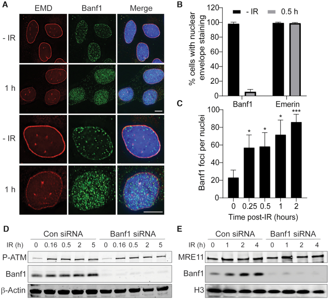
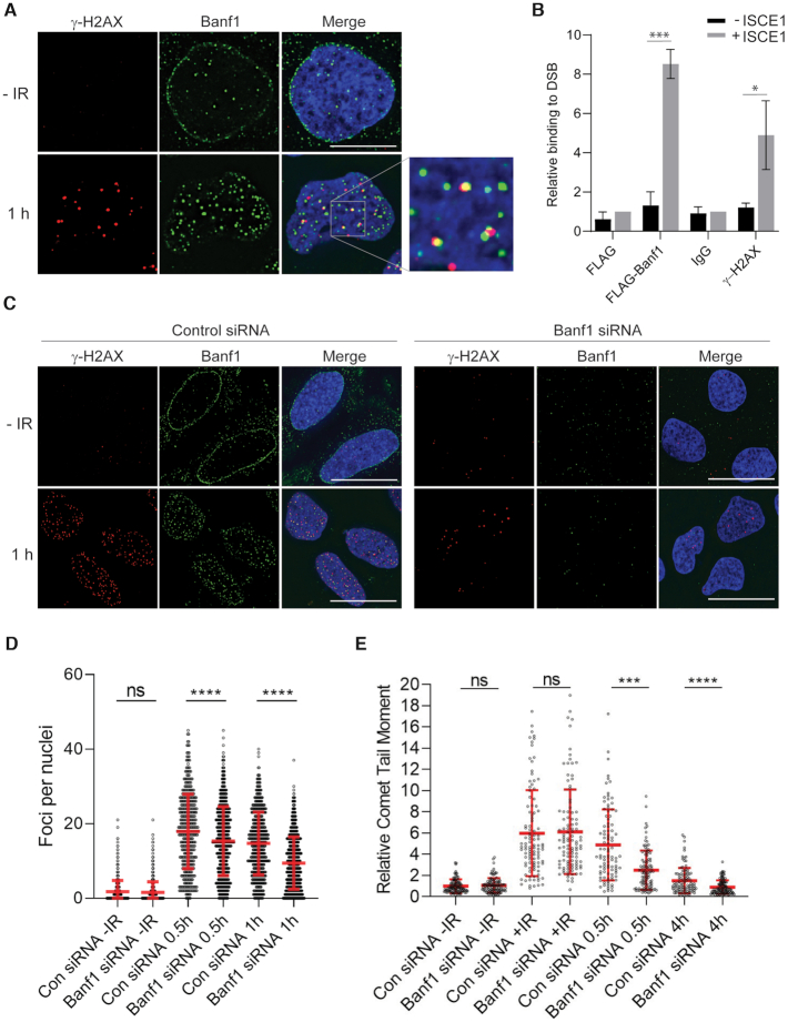
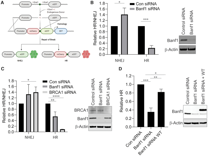
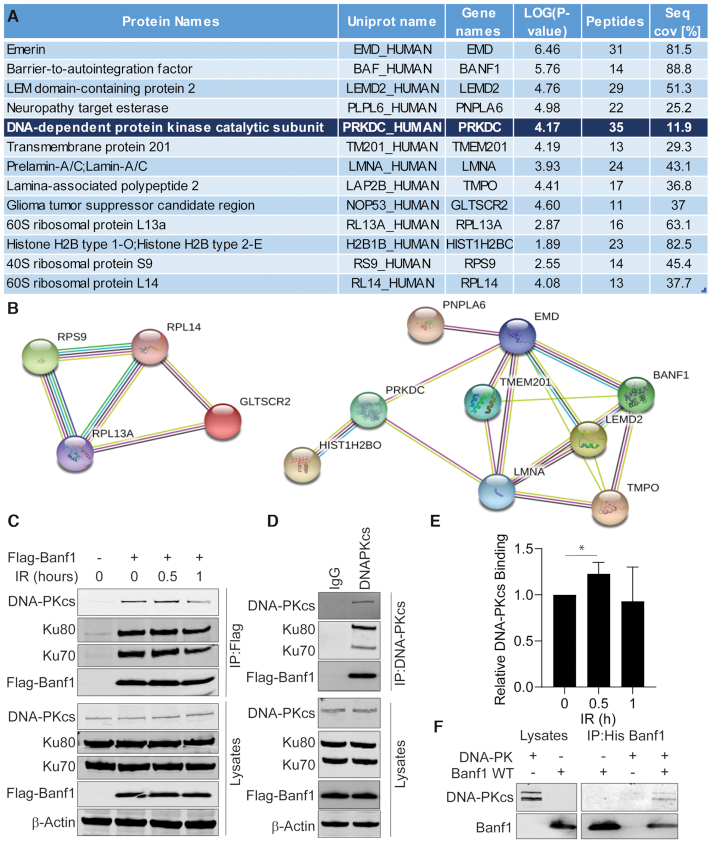
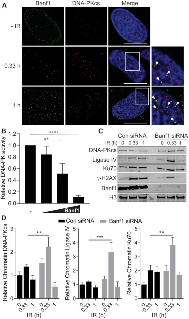
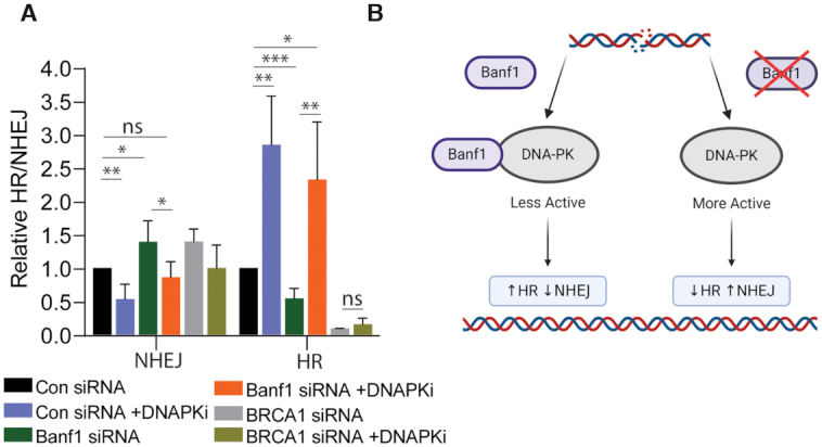
DNA repair pathways are essential to maintain the integrity of the genome and prevent cell death and tumourigenesis. Here, we show that the Barrier-to-Autointegration Factor (Banf1) protein has a role in the repair of DNA double-strand breaks. Banf1 is characterized as a nuclear envelope protein and mutations in Banf1 are associated with the severe premature aging syndrome, Néstor-Guillermo Progeria Syndrome. We have previously shown that Banf1 directly regulates the activity of PARP1 in the repair of oxidative DNA lesions. Here, we show that Banf1 also has a role in modulating DNA double-strand break repair through regulation of the DNA-dependent Protein Kinase catalytic subunit, DNA-PKcs. Specifically, we demonstrate that Banf1 relocalizes from the nuclear envelope to sites of DNA double-strand breaks. We also show that Banf1 can bind to and directly inhibit the activity of DNA-PKcs. Supporting this, cellular depletion of Banf1 leads to an increase in non-homologous end-joining and a decrease in homologous recombination, which our data suggest is likely due to unrestrained DNA-PKcs activity. Overall, this study identifies how Banf1 regulates double-strand break repair pathway choice by modulating DNA-PKcs activity to control genome stability within the cell.


Figures

Figure 1.
Figure 1.
Banf1 responds to ionizing radiation. (A) Banf1 relocalizes from the nuclear envelope following ionizing radiation in U2OS cells. Representative cells stained with the indicated antibodies at the indicated times post 6 Gy IR are shown. (B) Nuclear envelope localization of Banf1 (from a) was manually quantified using images taken on a Delta Vision PDV microscope. (C) Banf1 forms nuclear foci following IR. The number of Banf1 foci in U2OS cells treated as in a, were analysed via an InCell Analyser. (D) Banf1 protein levels stabilize following IR treatment. U2OS cells were transfected with control or Banf1 siRNA, treated with 6 Gy IR and whole cell lysates prepared at the indicated times post-IR. Lysates were immunoblotted with the indicated antibodies. P-ATM S1981 was used as a marker for DNA damage induction. (E) Banf1 is recruited to chromatin. U2OS cells were transfected with control or Banf1 siRNA, treated with 6 Gy IR and cells processed for cellular fractionation at the indicated times post-IR. The chromatin fraction was immunoblotted with the indicated antibodies. The accumulation of MRE11 was used as a marker for DNA damage and chromatin. Immunofluorescence scale bars represents 10 μm. Histogram data shown represent the mean and S.D. of three independent experiments and t-test was used for statistical analysis *P<0.05, ***P <0.001.
Figure 2.
Figure 2.
Banf1 localizes to DNA double-strand breaks. (A) Representative cells stained with the indicated antibodies at the indicated times post 2 Gy IR are shown. (B) ChIP analysis of Banf1 on a unique DSB induced by I-Sce1. Quantitative PCR was performed with primers at 94–378 bp from the break site in MCF7 DRGFP cells transfected with FLAG and Banf1-FLAG, ± I-Sce1. The enrichment of Banf1 after induction of a DSB was compared to the FLAG + I-Sce1 sample. γ-H2AX was also included as a control. (C) γ-H2AX foci in Banf1-depleted cells. U2OS cells were transfected with control or Banf1 siRNA. Seventy-two hours after transfection cells were treated with 2 Gy IR and fixed and stained with the indicated antibodies at the indicated time points. Representative cells are shown. (D) Cells from c were analysed for the number of γ-H2AX foci per cell using a Incell Analyser at the indicated times post-IR. Data points represent the γ-H2AX foci/nuclei from a minimum of 500 nuclei. Data shown show the mean and S.D. and are representative of three independent experiments. (E) Depletion of Banf1 promotes repair of IR-induced DNA damage. Neutral comet assay showing the relative Olive tail moment in U2OS control cells and cells depleted of Banf1 with siRNA at the indicated time points post-IR. +IR represents immediately following IR. Data points represent the Olive tail moment from a minimum of 100 cells. Data shown show the mean and S.D. of two independent experiments. Unless otherwise stated, data shown represent the mean and S.D. of three independent experiments and t-test was used for statistical analysis: ***P <0.001, ****P <0.0001. Immunofluorescence scale bars represent 10 μm.
Figure 3.
Figure 3.
Banf1 depletion inhibits HR and promotes NHEJ. (A) Schematic showing the possible outcomes of the HR/NHEJ reporter assay. (B) Depletion of Banf1 results in decreased HR and increased NHEJ. U2OS cells stably expressing a HR/NHEJ reporter were depleted of Banf1 using siRNA. Cells were transfected with an Isce1-expressing plasmid and a donor plasmid and GFP and mCherry positive cells were detected via FACS. (C) Depletion of Banf1 using esiRNA results in decreased HR and increased NHEJ. U2OS cells stably expressing a HR/NHEJ reporter were depleted of the indicated proteins using esiRNA. Cells were then transfected with an Isce1-expressing plasmid and a donor plasmid and GFP and mCherry positive cells were detected via FACS. (D) Restoration of Banf1 restores HR activity. U2OS cells stably expressing a HR/NHEJ reporter were depleted of Banf1 using siRNA. Cells were then transfected with empty Flag or siRNA resistant Flag-Banf1, an Isce1-expressing plasmid and a donor plasmid and GFP and mCherry positive cells were detected via FACS. Unless otherwise stated, data shown represent the mean and S.D. of 3 independent experiments and t-test was used for statistical analysis: *P < 0.05 **P < 0.01, ***P < 0.001, ****P < 0.0001.
Figure 4.
Figure 4.
Banf1 interacts with DNA-Dependent Kinase (DNA-PK). (A) DNA-PK was identified as a Banf1 interacting protein via mass spectrometry. A list of significant Banf1-interacting proteins is shown, with a minimum cut-off of 12 peptides. (B) String interactions of significant proteins are shown. (C) Banf1 and DNA-PK are in a complex; Immunoprecipitations from HEK293T cells expressing Flag or Flag-Banf1 at the indicated times post-IR using Flag antibodies. Immunoprecipitates were immunoblotted with the indicated antibodies. (D) Immunoprecipitations from HEK293T cells expressing Flag-Banf1 using DNA-PK antibodies or IgG as a control. Immunoprecipitates were immunoblotted with the indicated antibodies. (E) The histogram represents the analysis of the relative Banf1 binding to DNA-PK at the indicated times post-IR via immunoprecipitation, from (C). (F) Banf1 and DNA-PK directly interact. The recombinant His-Banf1 protein was incubated with purified DNA-PK complex (comprising of DNA-PKcs and Ku70/80) before immunoprecipitation with His beads and immunoblotting with the indicated antibodies. Unless otherwise stated, data shown represent the mean and S.D. of three independent experiments and unpaired t-test was used for statistical analysis: *P < 0.05.
Figure 5.
Figure 5.
Banf1 inhibits the kinase activity of DNA-PKcs. (A) Banf1 and DNA-PKcs partially colocalize following ionizing radiation. U2OS cells were treated or mock-treated with 6 Gy IR and treated with extraction buffer (+ RNase) prior to fixation in PFA at the indicated timepoints. Cells were then stained with the indicated antibodies. (B) Recombinant WT Banf1-mediated inhibition of the kinase activity of purified DNA-PK complex (comprising of DNA-PKcs and Ku70/80) on an immobilized substrate peptide. (C) Depletion of Banf1 leads to increased recruitment of NHEJ proteins to chromatin. U2OS cells were transfected with control or Banf1 siRNA. Cells were treated or mocked treated with 6 Gy IR and cellular fractionations processed at the indicated time post-treatment. Cellular fractions were immunoblotted with the indicated antibodies. Data shown are representative of four independent experiments. (D) The bands of DNA-PKcs, Ligase IV and Ku70 were analysed via densitometry and normalized to Histone H3 bands. Immunofluorescence scale bars represent 10 μm. Histogram data shown represent the mean and S.D. of four independent experiments. Unpaired t-test was used for statistical analysis. Unless otherwise stated, histogram data shown represent the mean and S.D. of three independent experiments, t-test was used for statistical analysis: **P < 0.01, ***P < 0.001, ****P < 0.0001.
Figure 6.
Figure 6.
Inhibition of DNA-PKcs activity restores HR activity in Banf1-depleted cells. (A) Inhibition of DNA-PKcs can restore HR in Banf1-depleted cells. U2OS cells stably expressing a HR/NHEJ reporter were depleted of the indicated proteins using esiRNA. Cells were then treated or mocked treated with a DNA-PKi, transfected with an Isce1-expressing plasmid and a donor plasmid and GFP and mCherry positive cells were detected via FACS. (B) Model of Banf1-mediated control of DSB repair pathway choice. In the presence of Banf1, DNA-PKcs is less active, leading to HR. Conversely, when Banf1 is depleted DNA-PKcs is more active promoting NHEJ. Image was created using BioRender.com. Histogram data shown represent the mean and S.D. of three independent experiments, t-test was used for statistical analysis: *P < 0.05 **P < 0.01, ***P < 0.001.

Similar articles

Cited by

References

    1. Ciccia A., Elledge S.J.. The DNA damage response: making it safe to play with knives. Mol. Cell. 2010; 40:179–204. - PMC - PubMed
    1. Ceccaldi R., Rondinelli B., D’Andrea A.D.. Repair pathway choices and consequences at the Double-Strand break. Trends Cell Biol. 2016; 26:52–64. - PMC - PubMed
    1. Jette N., Lees-Miller S.P.. The DNA-dependent protein kinase: a multifunctional protein kinase with roles in DNA double strand break repair and mitosis. Prog. Biophys. Mol. Biol. 2015; 117:194–205. - PMC - PubMed
    1. Xu Y., Xu D.. Repair pathway choice for double-strand breaks. Essays Biochem. 2020; 64:765–777. - PubMed
    1. Segura-Totten M., Kowalski A.K., Craigie R., Wilson K.L.. Barrier-to-autointegration factor: major roles in chromatin decondensation and nuclear assembly. J. Cell Biol. 2002; 158:475–485. - PMC - PubMed

Publication types

Substances