. 2021 Dec;20(6):836-852.
doi: 10.1007/s12311-021-01246-7. Epub 2021 Mar 4.

Functional Convergence of Motor and Social Processes in Lobule IV/V of the Mouse Cerebellum

Affiliations

Functional Convergence of Motor and Social Processes in Lobule IV/V of the Mouse Cerebellum

Owen Y Chao et al. Cerebellum. 2021 Dec.

Abstract

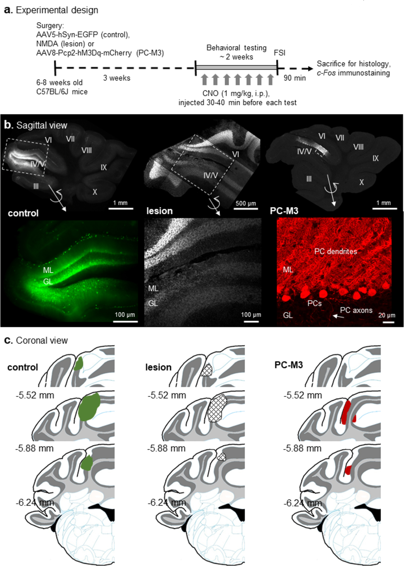
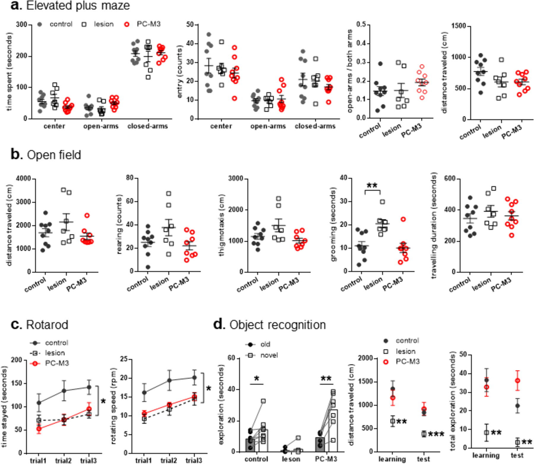
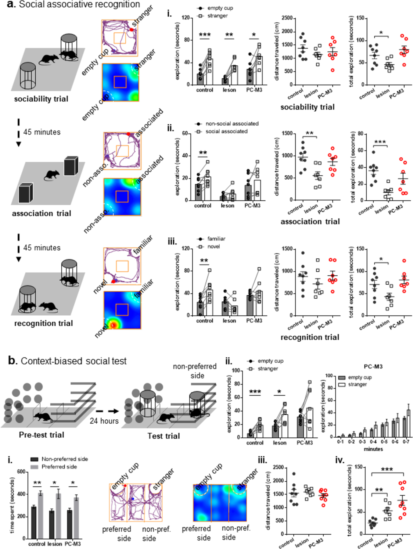
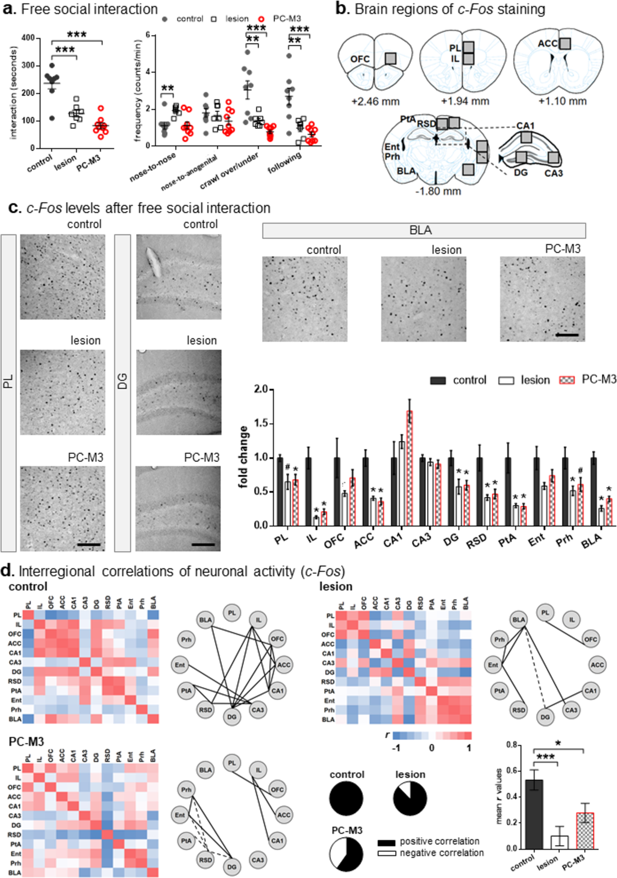
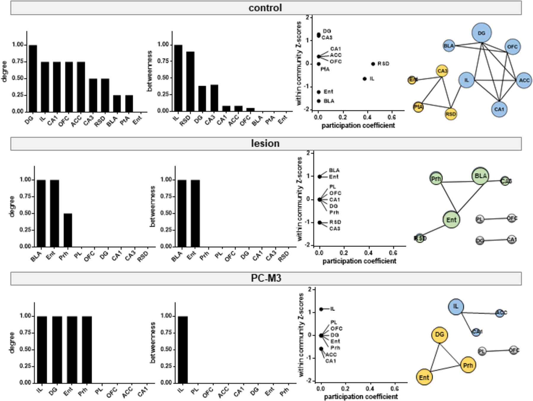
Topographic organization of the cerebellum is largely segregated into the anterior and posterior lobes that represent its "motor" and "non-motor" functions, respectively. Although patients with damage to the anterior cerebellum often exhibit motor deficits, it remains unclear whether and how such an injury affects cognitive and social behaviors. To address this, we perturbed the activity of major anterior lobule IV/V in mice by either neurotoxic lesion or chemogenetic excitation of Purkinje cells in the cerebellar cortex. We found that both of the manipulations impaired motor coordination, but not general locomotion or anxiety-related behavior. The lesioned animals showed memory deficits in object recognition and social-associative recognition tests, which were confounded by a lack of exploration. Chemogenetic excitation of Purkinje cells disrupted the animals' social approach in a less-preferred context and social memory, without affecting their overall exploration and object-based memory. In a free social interaction test, the two groups exhibited less interaction with a stranger conspecific. Subsequent c-Fos imaging indicated that decreased neuronal activities in the medial prefrontal cortex, hippocampal dentate gyrus, parahippocampal cortices, and basolateral amygdala, as well as disorganized modular structures of the brain networks might underlie the reduced social interaction. These findings suggest that the anterior cerebellum plays an intricate role in processing motor, cognitive, and social functions.

Keywords: Cerebellum; Chemogenetics; Functional connectivity; Lesion; Object recognition; Social memory.


Conflict of interest statement

Conflict of interests

The authors declare no competing financial and non-financial interests.

Figures

Fig 1.
Fig 1.. Experimental design and histological examination.
(a) Mice were randomly divided into AAV5-hSyn-EGFP (control), NMDA (lesion) and AAV8-Pcp2-hM3Dq-mCherry (PC-M3) groups, followed by a series of behavioral tests. Clozapine-N-oxide (CNO, 1 mg/kg) was given to all the groups before each test. Mice were sacrificed 90 min after the final test (free social interaction, FSI) and c-Fos immunostaining was performed. (b) Examples of the sagittal view of cerebellar slices from the control, lesion and PC-M3 groups. Fluorescence in lobule IV/V was EGFP (control), DAPI (lesion) or mCherry (PC-M3). Zoom-in images are shown in bottom panels. In the control group, EGFP was found preferentially in granule cells and interneurons. In the lesion group, cell loss was seen in molecular layer (ML), Purkinje cell (PC) layer, and granular layer (GL). In the PC-M3 group, only PCs were labelled with mCherry. (c) Diagrams of the coronal view of cerebellar sections. Affected areas in lobule IV/V are highlighted in green (EGFP in control group), grid-shaped (lesions in lesion group) and red (mCherry in PC-M3 group), respectively. Numbers indicate distance relative to the bregma.
Fig 2.
Fig 2.. Effects of lesion or chemogenetic excitation of PCs in lobule IV/V on anxiety-related behavior, locomotor activity, motor coordination and object memory.
(a) No significant differences were found between control and lesioned or PC-M3 animals in the elevated plus maze. (b) In the open field, the lesion group had more self-grooming than the controls, while both lesion and PC-M3 groups showed comparable locomotor activities to the controls. (c) The lesion and PC-M3 groups displayed impaired motor coordination on an accelerating rotarod. (d) The control and PC-M3 groups showed intact object memory, but the lesion group did not. The lesioned animals also exhibited less motor activity and exploration of the objects than the controls. n=7–9/group. Values are shown as mean ± SEM. *p<0.05, **p<0.01, ***p<0.001, compared to the control group or respective values.
Fig 3.
Fig 3.. Lesion or chemogenetic manipulation in lobule IV/V disrupted social behaviors.
(a) Illustration of the social associative recognition test, including three trials: sociability (exploration of a stranger and an empty cup), association (exploration of two identical objects, placed at the same locations of the previously explored stimuli) and recognition (exploration of the previously encountered stranger and a novel stranger). Examples of movement tracking (blue and red dots indicate the start and end points, respectively) and heat-map of time spent (the spectra from red to blue represents long to short duration) from a control animal were presented. (i) All the groups explored the stranger more than the empty cup in the sociability trial, while the lesion group had less total exploration. (ii) Only the control group explored the associated object more than the non-associated object in the association trial. The lesioned animals had less distanced travelled and total exploration than the controls. (iii) Only the control group explored the novel stranger more than the familiar one. The lesion group again showed less total exploration than the control group. (b) Diagram of the context-biased social test: exploration of the three-chambered box with two distinct contexts (pre-test trial), followed by exploration of a stranger located in the non-preferred side and an empty cup located in the other side (test trial). (i) All the groups formed a natural contextual preference in the pre-test trial. An example of movement tracking and heat-map of time spent from a control animal in the test trial was presented. (ii) In the test trial, the control and lesion groups explored the stranger located in the non-preferred context more than the empty cup located in the preferred context, while the PC-M3 group did not across time periods of the test. (iii) No group difference was found in distance travelled in the test trial. (iv) Yet, the lesion and PC-M3 groups had higher total exploration than the control group. n=7–8/group. Values are shown as mean ± SEM. *p<0.05, **p<0.01, ***p<0.001, compared to the control group or respective values.
Fig 4.
Fig 4.. Lesion or chemogenetic manipulation in lobule IV/V reduced free social interaction and c-Fos levels in multiple brain regions.
(a) The lesion and PC-M3 groups displayed less social interaction and lower frequencies of crawling and following behaviors than the control group. The lesioned mice had more nose-to-nose/head contacts than the controls. n=7–9/group. (b) Brain regions imaged for c-Fos-positive cells. Numbers indicate mean distance relative to the bregma in ± 0.1–0.2 mm range. (c) Examples of c-Fos images in the PL, DG and BLA, and fold change of c-Fos-positive cells measured across the brain regions. Scale bars: 50 μm. The lesion and PC-M3 groups showed fewer c-Fos signals in the PL, IL, ACC, DG, RSD, PtA, Prh and BLA than the control group. n=5–6/group. (d) Matrices of interregional correlations for c-Fos-positive cells and significant connections (solid and dash lines represent positive and negative correlations, respectively) in the three groups. Relative proportions of positive and negative correlations for each group are shown in the pie charts. The lesion and PC-M3 groups had lower average correlations r than the control group in the calculation of all interregional correlation coefficients. Values are shown as mean ± SEM. *p<0.05, **p<0.01, ***p<0.001, #p=0.052, compared to the control group. PL: prelimbic cortex; IL: infralimbic cortex; OFC: orbitofrontal cortex; ACC: anterior cingulate cortex; DG: dentate gyrus; RSD: dorsal retrosplenial cortex; PtA: parietal association cortex; Ent: entorhinal cortex; Prh: perirhinal cortex; BLA: basolateral amygdala.
Fig 5.
Fig 5.. Lesion or chemogenetic perturbation of lobule IV/V disorganized neural networks required for free social interaction.
Brain hubs identified by graph theoretical analysis of the networks activated following free social interaction. Normalized degree and betweenness are ranked in descending order for each group. Neuronal modules are identified via modularity maximization, based on within-community Z-scores and participation coefficients for each brain region. Different modules are color-coded and the size of nodes (brain regions) is proportional to their degree in the network. PL: prelimbic cortex; IL: infralimbic cortex; OFC: orbitofrontal cortex; ACC: anterior cingulate cortex; DG: dentate gyrus; RSD: dorsal retrosplenial cortex; PtA: parietal association cortex; Ent: entorhinal cortex; Prh: perirhinal cortex; BLA: basolateral amygdala.

References

    1. Purves D, et al., Neuroscience. 2019.
    1. Koziol LF, et al., Consensus paper: the cerebellum’s role in movement and cognition. Cerebellum, 2014. 13(1): p. 151–77. - PMC - PubMed
    1. Sokolov AA, Miall RC, and Ivry RB, The Cerebellum: Adaptive Prediction for Movement and Cognition. Trends Cogn Sci, 2017. 21(5): p. 313–332. - PMC - PubMed
    1. Sathyanesan A, et al., Emerging connections between cerebellar development, behaviour and complex brain disorders. Nature Reviews Neuroscience, 2019. 20(5): p. 298–313. - PMC - PubMed
    1. Schmahmann JD, et al., The Theory and Neuroscience of Cerebellar Cognition. Annu Rev Neurosci, 2019. 42: p. 337–364. - PubMed

LinkOut - more resources