. 2021 Mar 4;16(3):e0247752.
doi: 10.1371/journal.pone.0247752. eCollection 2021.

Isoalantolactone inhibits pancreatic cancer proliferation by regulation of PI3K and Wnt signal pathway

Affiliations

Isoalantolactone inhibits pancreatic cancer proliferation by regulation of PI3K and Wnt signal pathway

Chaoxiong Zhang et al. PLoS One. .

Abstract

Background/aims: Isoalantolactone (IATL) is one of multiple isomeric sesquiterpene lactones and is isolated from inula helenium. IATL has multiple functions such as antibacterial, antihelminthic and antiproliferative activities. IATL also inhibits pancreatic cancer proliferation and induces apoptosis by increasing ROS production. However, the detailed mechanism of IATL-mediated pancreatic cancer apoptosis remains largely unknown.

Methods: In current study, pancreatic carcinoma cell lines (PANC-1, AsPC-1, BxPC-3) and a mouse xenograft model were used to determine the mechanism of IATL-mediated toxic effects.

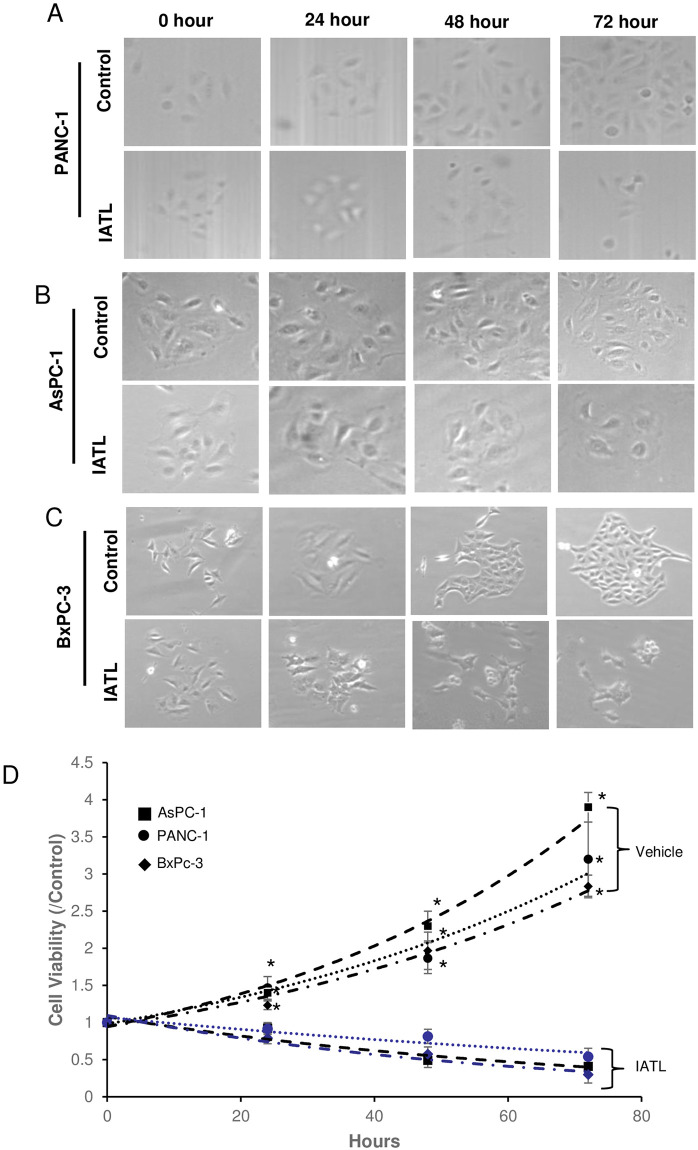
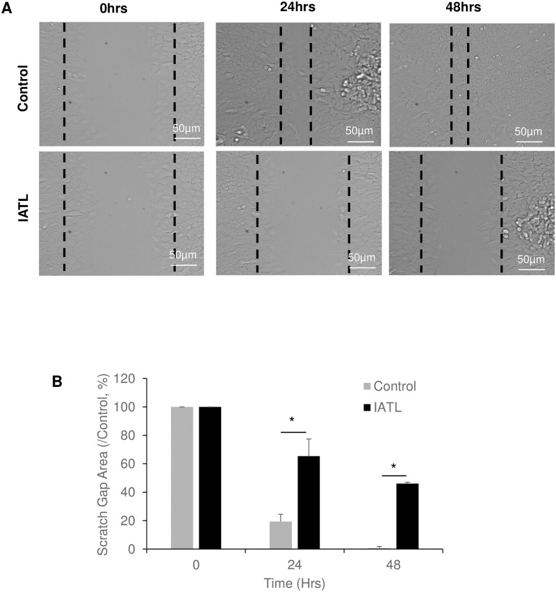
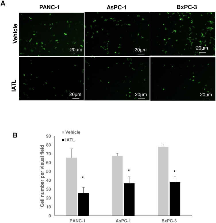
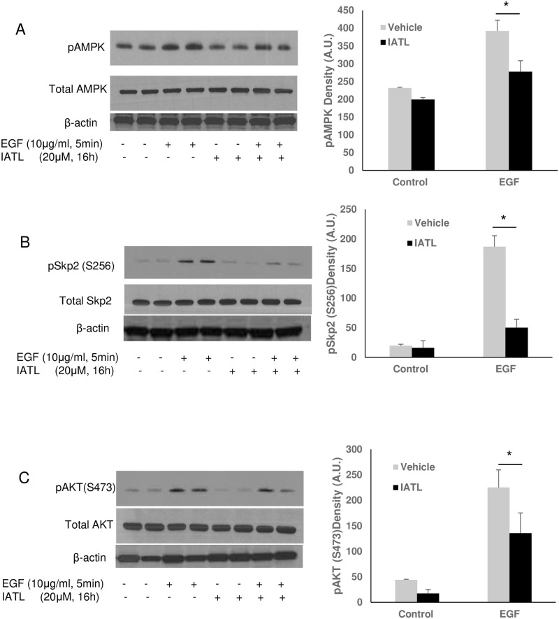
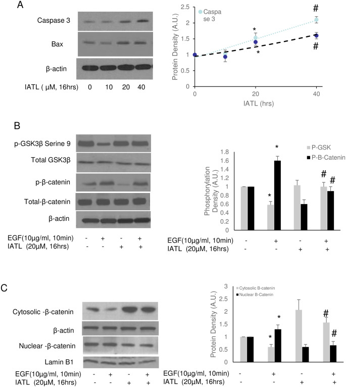
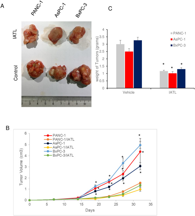
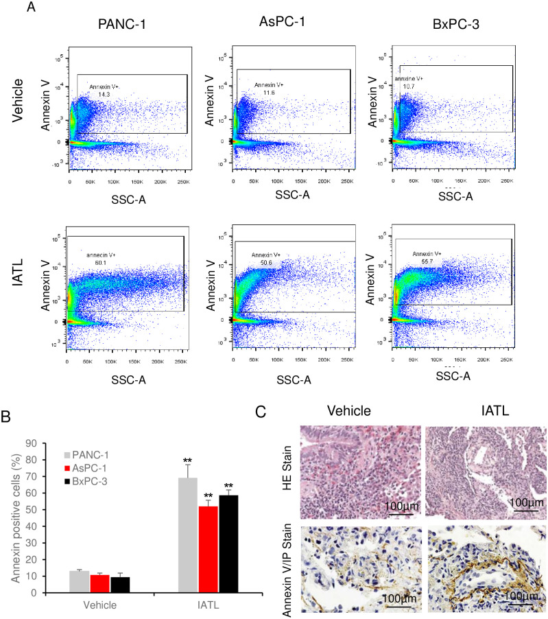
Results: IATL (20μM) inhibited pancreatic adenocarcinoma cell lines proliferation in a time-dependent way; while scratch assay showed that IATL significantly inhibited PANC-1 scratch closure (P<0.05); Invasion assays indicated that IATL significantly attenuated pancreatic adenocarcinoma cell lines invasion on matrigel. Signal analysis showed that IATL inhibited pancreatic adenocarcinoma cell proliferation by blocking EGF-PI3K-Skp2-Akt signal axis. Moreover, IATL induced pancreatic adenocarcinoma cell apoptosis by increasing cytosolic Caspase3 and Box expression. This apoptosis was mediated by inhibition of canonical wnt signal pathway. Finally, xenograft studies showed that IATL also significantly inhibited pancreatic adenocarcinoma cell proliferation and induced pancreatic adenocarcinoma cell apoptosis in vivo.

Conclusions: IATL inhibits pancreatic cancer proliferation and induces apoptosis on cellular and in vivo models. Signal pathway studies reveal that EGF-PI3K-Skp2-Akt signal axis and canonical wnt pathway are involved in IATL-mediated cellular proliferation inhibition and apoptosis. These studies indicate that IATL may provide a future potential therapy for pancreatic cancer.


Conflict of interest statement

The authors have declared that no competing interests exist.

Figures

Fig 1
Fig 1. IATL inhibits pancreatic cancer cell proliferation.
The PANC-1, AsPC-1 or BxPC-3 cells were treated with 20μM IATL at the indicated times. After the cells were digested with trypsin, the cellular viability was determined by MTT assay. (A) representative images of PANC-1 were treated with IATL; (B) The morphological characteristic changes in AsPC-1 when treated with IATL; (C) The morphological characteristic changes in BxPc-3 when treated with IATL; (D) MTT assay indicated that IATL significantly inhibited cell viability in each cell line (*P<0.05, n = 4).
Fig 2
Fig 2. IATL inhibits pancreatic cancer cell migration.
The confluent PANC-1 cells were scratched followed by treatment with 20 μM IATL at the indicated times. (A) Representative scratch images of PANC-1 in the presence or absence of IATL; (B) IATL significantly inhibited scratch closure of PANC-1 as compared to vehicle treatment. The PANC cell closure areas were represented as percentage of 0 hours, respectively (P<0.05, n = 3).
Fig 3
Fig 3. IATL inhibits pancreatic cancer cell invasion.
The GFP-expressing PANC-1, AsPC-1 or BxPC-3 cells were seeded into transwell inserts, which were previously coated with matrigel. The PANC-1, AsPC-1 or BxPC-3 cells (1×10⁵) were treated with 20 μM IATL or vehicle for 24hours. The insert membranes were cut and mounted onto coverslides and the images were taken using fluorescent microscopy. (A) Representative cell images on the insert member after IATL or vehicle treatment for 24 hours; (B) Compared to vehicle, IATL treatment significantly inhibited invasion in all three different pancreatic cancer cell lines (P<0.05, n = 4).
Fig 4
Fig 4. IATL inhibits EGF signal pathway through Skp2 inactivation in pancreatic cancer cells.
The PANC-1 cells were treated with 20μM IATL followed by EGF (10μg/mL) treatment for 5minutes. After the cell lysates were harvested, the target protein phosphorylation was determined by western blotting. EGF caused AMPK (A), Skp2 (B) and Akt (C) phosphorylation increases; but IATL significantly inhibited EGF-mediated EGF (A)-SKp2 (B) and Akt (C) phosphorylation (P<0.05, n = 3).
Fig 5
Fig 5. IATL inhibits Wnt signal pathway and induces pancreatic cancer cells apoptosis.
The PANC-1 cells were treated with 20μM IATL alone or in combination with EGF (5μg/mL) at the indicated time. The cell lysates were harvested for western blotting. (A) IATL induced both Caspase 3 and Bax in PANC-1 cells in a dose-dependent fashion; (B) EGF activated the Wnt signal pathway by decreasing serine 9 phosphorylation of GSK-3β (*P<0.05, n = 3), however, IATL increased GSK-3β phosphorylation (#P<0.05, n = 3); EGF increased β-catenin phosphorylation (*P<0.05) but IATL reversed this regulation (#P<0.05, n = 3); (C) EGF caused β-catenin translocation into cellular nucleus (*P<0.05, n = 3) but IATL inhibited EGF-mediated β-catenin translocation (#P<0.05, n = 3).
Fig 6
Fig 6. IATL inhibits pancreatic cancer cells growth in vivo.
The PANC-1, AsPC-1 or BxPC-3 cells were subcutaneously xenografted onto BALB/c nude mice flank. Once the cells were injected, the mice were treated with IATL (0.5mg/kg) or vehicle once a week for 5 weeks. At the end of experiment, the xenografts were removed for assessing weight or for imaging. (A) Images of representative tumors from PANC-1, AsPC-1 or BxPC-3 cell xenografts (n = 6). (B) Growth curve of tumors xenografts in the presence or absence of IATL over 5 weeks; (C) IATL significantly decreased the weight of tumors generated from PANC-1, AsPC-1 or BxPC-3 cells in vivo (*, P<0.05).
Fig 7
Fig 7. IATL induces pancreatic cancer cells apoptosis in vivo.
The PANC-1, AsPC-1 or BxPC-3 cells were xenografted on BALB/c nude mice and were treated with IATL. In the end of experiment, the xenografts on nude mice were removed for flowcytometry or histology assay. (A) Representative images of flow cytometry assay showing detection of annexin-V positive cells from PANC-1, AsPC-1 or BxPC-3 xenografts for both vehicle and IATL treatment groups; (B) Compared to vehicle, IATL treatment significantly increased annexin-V positive cells in PANC-1, AsPC-1 or BxPC-3 cell xenografts (*, P<0.05; **, <0.01). (C) Representative images of H&E staining (upper panel) and annexin V staining (bottom panel) of PANC-1s xenografts (n = 6).

References

    1. Collisson EA, Bailey P, Chang DK, Biankin AV. Molecular subtypes of pancreatic cancer. Nat Rev Gastroenterol Hepatol. 2019;16(4):207–20. 10.1038/s41575-019-0109-y - DOI - PubMed
    1. Kota J, Hancock J, Kwon J, Korc M. Pancreatic cancer: Stroma and its current and emerging targeted therapies. Cancer Lett. 2017;391:38–49. 10.1016/j.canlet.2016.12.035 - DOI - PubMed
    1. Rawla P, Thandra KC, Sunkara T. Pancreatic cancer and obesity: epidemiology, mechanism, and preventive strategies. Clin J Gastroenterol. 2019. - PubMed
    1. Garcia-Reyes B, Kretz AL, Ruff JP, von Karstedt S, Hillenbrand A, Knippschild U, et al.. The Emerging Role of Cyclin-Dependent Kinases (CDKs) in Pancreatic Ductal Adenocarcinoma. Int J Mol Sci. 2018;19(10). - PMC - PubMed
    1. Song M, Bode AM, Dong Z, Lee MH. AKT as a Therapeutic Target for Cancer. Cancer Res. 2019;79(6):1019–31. 10.1158/0008-5472.CAN-18-2738 - DOI - PubMed

MeSH terms