. 2021 Mar 4;16(3):e0248027.
doi: 10.1371/journal.pone.0248027. eCollection 2021.

Polymorphic SERPINA3 prolongs oligomeric state of amyloid beta

Affiliations

Polymorphic SERPINA3 prolongs oligomeric state of amyloid beta

Maruf Mohammad Akbor et al. PLoS One. .

Abstract

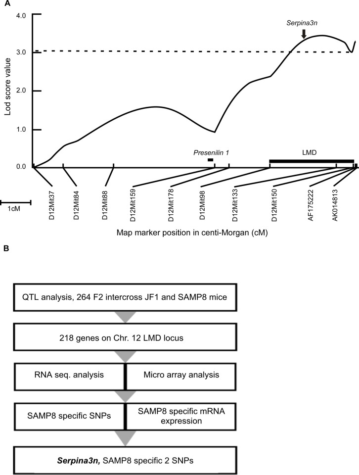
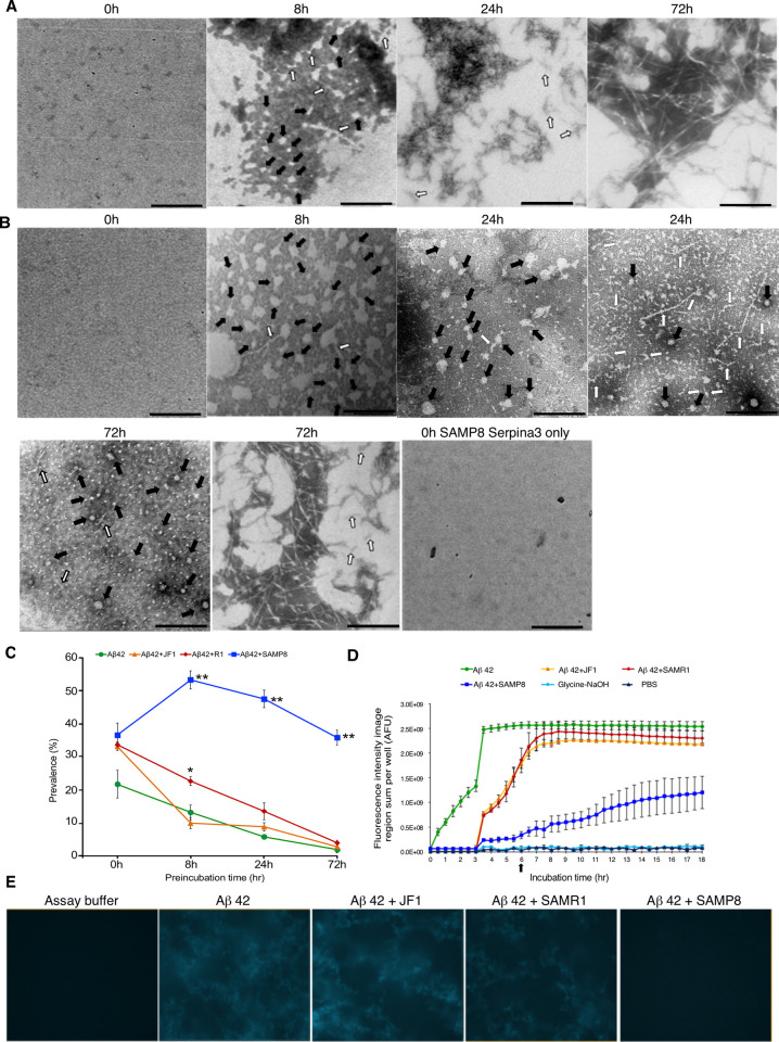
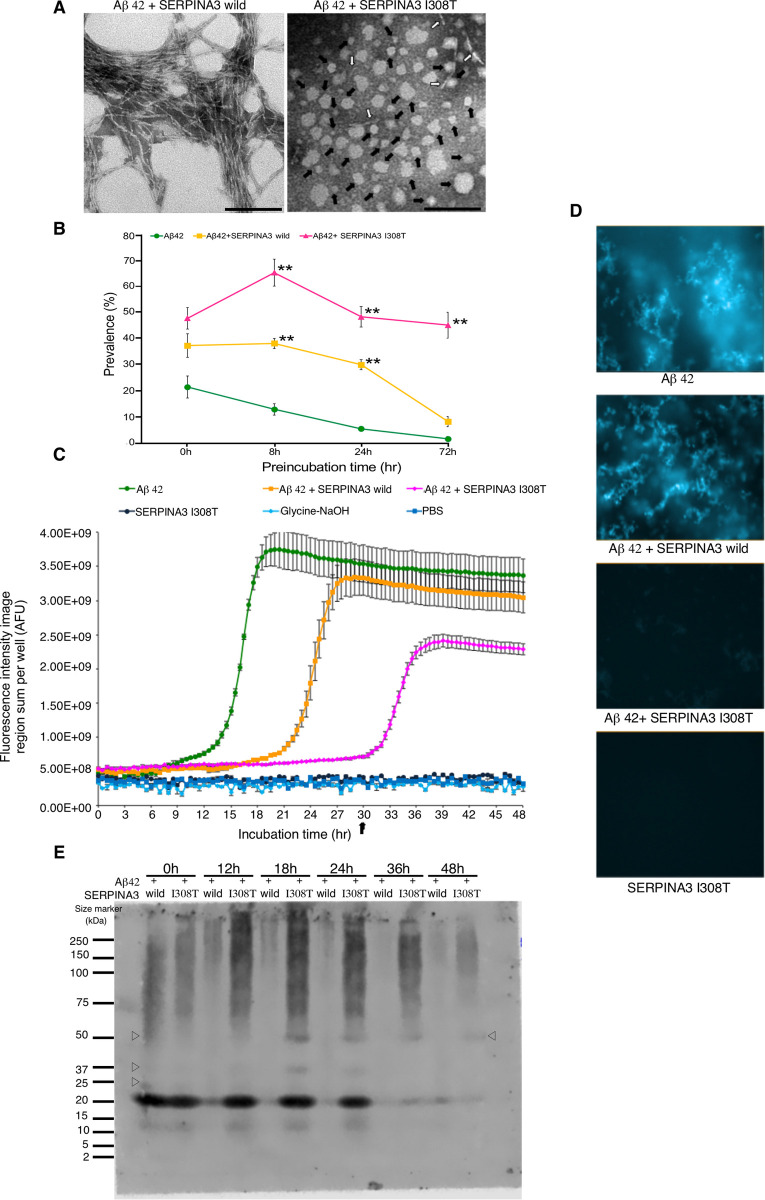
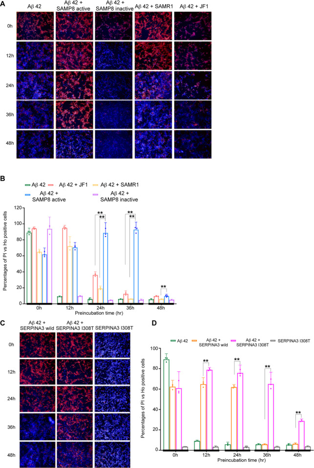
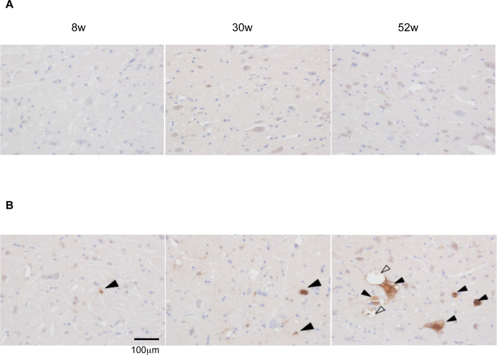
Molecular chaperon SERPINA3 colocalizes with accumulated amyloid peptide in Alzheimer's disease (AD) patient's brain. From the QTL analysis, we narrowed down Serpina3 with two SNPs in senescence-accelerated mouse prone (SAMP) 8 strain. Our study showed SAMP8 type Serpina3 prolonged retention of oligomeric Aβ 42 for longer duration (72 hr) while observing under transmission electron microscope (TEM). From Western blot results, we confirmed presence of Aβ 42 oligomeric forms (trimers, tetramers) were maintained for longer duration only in the presences of SAMP8 type Serpina3. Using SH-SY5Y neuroblastoma cell line, we observed until 36 hr preincubated Aβ 42 with SAMP8 type Serpina3 caused neuronal cell death compared to 12 hr preincubated Aβ 42 with SAMR1 or JF1 type Serpina3 proteins. Similar results were found by extending this study to analyze the effect of polymorphism of SERPINA3 gene of the Japanese SNP database for geriatric research (JG-SNP). We observed that polymorphic SERPINA3 I308T (rs142398813) prolonged toxic oligomeric Aβ 42 forms till 48 hr in comparison to the presence wild type SERPINA3 protein, resulting neuronal cell death. From this study, we first clarified pathogenic regulatory role of polymorphic SERPINA3 in neurodegeneration.


Conflict of interest statement

The authors have declared that no competing interests exist.

Figures

Fig 1
Fig 1. QTL analysis to identify candidate gene on chromosome 12 LMD locus.
(A) Lod (logarithm of odd) score plots for LMD phenotype on chromosomes 12. X-axis: Indicates map position in centi-Morgan (cM). Microsatellite markers are indicated. Y-axis: Lod scores for QTL in (SAMP8 x JF1) F2 progeny. Vertical dotted line on lod plot indicates the lod = 3.1 significance threshold. Both male and female scores were standardized and analyzed by Mapmaker/QTL. Thick small horizontal line indicates the 1-lod support interval for the noted LMD locus. Solid arrow indicates position of Serpina3n (104,406,729–104,414,329 bp), vertical bar indicates position of Presenilin 1 (83,688,152–83,735,199 bp). (B) Strategy to narrow down SAMP8 specific SNPs.
Fig 2
Fig 2. Regulation of various Aβ peptide forms by mouse polymorphic Serpina3n.
Representative TEM images of Aβ peptide at different preincubation time points using (A) Aβ 42 peptide. (B) Aβ 42 + SAMP8 Serpina3n. 0 hr preincubation did not show any define structure either alone or in presence of both Aβ peptide with SAMP8 Serpina3n protein. Solid arrow indicated oligomeric peptide, while open arrow indicated protofibrillar form of Aβ peptide. Representative fibrillar form of Aβ peptides were shown at 24 hr (Fig 2A) and 72 hr (Fig 2A and 2B) of preincubation. Scale bar: 100 nm. (C) Kinetics of amyloid β peptide oligomerization under influence of polymorphic mouse Serpina3n. Here, x-axis shows prevalence of oligomer rich observation fields (in percentage) and y-axis shows preincubation time (hr). Significance levels were tested SAMP8 vs. SAMR1; SAMP8 vs. JF1; SAMR1 vs. JF1. (n = 3, * P ≤ 0.05; ** P ≤ 0.01. One-way ANOVA with Tukey and Kramer’s honestly significance difference test was used). (D) Thioflavin T fluorescence assay (data represent Mean ± SEM, n = 3). AFU refers arbitrary fluorescence units. (E) Representative images of thioflavin T fluorescence assay at 6 hrs.
Fig 3
Fig 3. Regulation of various Aβ peptide forms by human polymorphic SERPINA3.
(A) Representative TEM images of Aβ peptide at 72 hr preincubation. Typical fibrillar form was observed in Aβ 42 + SERPINA3 wild type; whereas Aβ 42 + SERPINA3 I308T still showed irregularly shaped oligomers as indicated by solid arrow and while open arrow indicated protofibrillar form. Scale bar: 100 nm. (B) Kinetics of amyloid β peptide oligomerization in presence of human SERPINA3. Here, x-axis shows prevalence of oligomer rich observation fields (in percentage) and y-axis shows preincubation time (hr). Significance levels were tested Aβ 42 + SERPINA3 I308T vs. Aβ 42 + SERPINA3 wild; Aβ 42 + SERPINA3 I308T vs. Aβ 42; Aβ 42 + SERPINA3 wild vs. Aβ 42. (n = 3, ** P ≤ 0.01. One-way ANOVA with Tukey and Kramer’s honestly significance difference test was used). (C) Thioflavin T fluorescence assay (data represent Mean ± SEM, n = 3). AFU refers arbitrary fluorescence units. (D) Representative images of thioflavin T fluorescence assay at 30 hrs. (E) Representative image of gradient gel native PAGE, showing Aβ 42 peptide in presence of human SERPINA3 recombinant proteins, at 0 to 48 hrs of preincubation. Open triangle showed presence of oligomeric Aβ peptide conformations. See also in S3 Fig.
Fig 4
Fig 4. Aβ 42 induced cytotoxicity using SHSY5Y neuroblastoma cell line.
Hoechst 33342 showing blue signal of intact nucleus; propidium iodide (PI) identified red signal of damaged cell DNA. (A) Representative images of cytotoxicity assay at different preincubation time points using mouse Serpina3n. (B) Bar graphs showing percentage ratio of PI vs. Hoechst 33342 positive cells in y-axis at different preincubated time points indicated with thick vertical line in x-axis. Significance levels were tested SAMP8 vs. SAMR1 & SAMP8 vs. JF1. (n = 3, * P ≤ 0.05; ** P ≤ 0.01. One-way ANOVA with Tukey and Kramer’s honestly significance difference test was used). (C) Representative images at different preincubation time points using human SERPINA3. (D) Bar graphs showing percentage ratio of PI vs. Hoechst 33342 positive cells. Significance levels were tested against human SERPINA3 I308T vs. SERPINA3 wild type. (n = 3, * P ≤ 0.05; ** P ≤ 0.01. Tukey and Kramer’s honestly significance difference test was used).
Fig 5
Fig 5. Expression of Serpina3 in mouse brain stem.
Representative brain slice images of different ages 8 weeks (left), 30 weeks (middle), 52 weeks (right) of SAMR1(upper panel) and SAMP8 mice (lower panel). (A) SAMR1 (B) SAMP8. In the brain stem of SAMP8, immunoreactivity for Serpina3 (solid arrow) was observed in some astrocytic and neuronal cells of SAMP8, started from 8 weeks followed by 30 and 52 weeks, especially in 52 weeks old SAMP8, while that was not observed in the brain stem of SAMR1. In the brain stem of 52-week-old SAMP8, immunoreactivity for Serpina3 (solid arrow) was observed in some neuronal cells showing vacuolar degeneration (open arrow) (scale bar = 100 μm).

Similar articles

Cited by

References

    1. Medway C, Morgan K. Review: The genetics of Alzheimer’s disease; putting flesh on the bones. Neuropathology and Applied Neurobiology. 2014; 40:97–05. 10.1111/nan.12101 - DOI - PMC - PubMed
    1. Kamboh MI, Demirci FY, Wang X, Minster RL, Carrasquillo MM, Pankratz VS, et al.. Genome-wide association study of Alzheimer’s disease. Translational Psychiatry. 2012; 2:e117. 10.1038/tp.2012.45 - DOI - PMC - PubMed
    1. Chen JA, Wang Q, Davis-Turak J, Li Y, Karydas AM, Hsu SC, et al.. A Multiancestral Genome-Wide Exome Array Study of Alzheimer Disease, Frontotemporal Dementia, and Progressive Supranuclear Palsy. JAMA Neurol. 2015; 72:414–22. 10.1001/jamaneurol.2014.4040 - DOI - PMC - PubMed
    1. Xia X, Jiang Q, McDermott J, Han JDJ. Aging and Alzheimer’s disease: Comparison and associations from molecular to system level. Aging Cell. 2018; 17:e12802. 10.1111/acel.12802 - DOI - PMC - PubMed
    1. Edwards GA III, Gamez N, Escobedo GJ, Calderon O, Moreno-Gonzalez I. Modifiable Risk Factors for Alzheimer’s Disease. Front. Aging Neurosci. 2019; 11:146. 10.3389/fnagi.2019.00146 - DOI - PMC - PubMed

Publication types

MeSH terms