18F-FDG PET/CT imaging for aggressive melanotic schwannoma of the L3 spinal root: A case report
- PMID: 33663098
- PMCID: PMC7909145
- DOI: 10.1097/MD.0000000000024803
18F-FDG PET/CT imaging for aggressive melanotic schwannoma of the L3 spinal root: A case report
Abstract
Rationale: Melanotic schwannoma (MS) is an unusual variant of a nerve sheath neoplasm that accounts for less than 1% of all primary peripheral nerve sheath tumors. Fluorine-18 fluorodeoxyglucose positron emission tomography/computed tomography (18F-FDG PET/CT) has unique value in detecting malignant MS lesions. To date, only 4 cases of MS with hepatic metastasis have been reported. Herein, we report the fifth case, which is the first reported patient with MS of Asian ethnicity with hepatic metastasis.
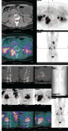
Patient concerns: A 29-year-old woman with a 1-day history of backache was admitted to our hospital. PET/CT showed a paravertebral heterogeneous soft tissue mass along the spinal nerve at the L2-L3 level with strong FDG uptake, and a nodule with increased FDG uptake in the lateral lobe of the left liver.
Diagnosis: A puncture biopsy of the L3 bony destruction and surrounding soft tissue mass was performed. The final diagnosis was spinal MS with hepatic metastasis.
Interventions: The patient underwent 6 courses of systemic chemotherapy.
Outcomes: The patient did not receive further treatment for half a year after the end of chemotherapy and recovered well.
Lessons: Unlike conventional schwannomas, which are completely benign, MS has an unpredictable prognosis. It is thought to have low malignant potential, and the malignant type tends to metastasize. FDG PET/CT has a unique and important value in the differential diagnosis of benign and malignant lesions, in detecting occult metastases, monitoring the treatment response, and assessing the prognosis of MS.
Copyright © 2021 the Author(s). Published by Wolters Kluwer Health, Inc.
Conflict of interest statement
The authors have no funding and conflicts of interest to disclose.
Figures


References
-
- Millar WG. A malignant melanotic tumor of ganglion cells arising from thoracic sympathetic ganglion. Pathol Bacteriol 1932;35:351–7.
-
- Solomon RA, Handler MS, Sedelli RV, et al. . Intramedullary melanotic schwannoma of cervicomedullary junction. Neurosurgery 1987;20:36–8. - PubMed
-
- Santaguida C, Sabbagh AJ, Guiot MC, et al. . Aggressive intramedullary melanotic schwannoma. Neurosurgery 2004;55:1430–4. - PubMed
-
- Zhang HY, Yang GH, Chen HJ, et al. . Clinicopathological, immunohistochemical, and ultrastructural study of 13 cases of melanotic schwannoma. Chin Med J 2005;118:1451–61. - PubMed
-
- Torres-Mora J, Dry S, Li X, et al. . Malignant melanotic schwannian tumor. A clinicopathologic, immunohistochemical, and gene expression profiling study of 40 cases, with a proposal for the reclassification of “melanotic schwannoma”. Am J Surg Pathol 2014;38:94–105. - PubMed
Publication types
MeSH terms
Substances
LinkOut - more resources
Full Text Sources
Other Literature Sources

