. 2021 Mar 4;22(1):153.
doi: 10.1186/s12864-021-07441-4.

A comparative analysis of heart microRNAs in vertebrates brings novel insights into the evolution of genetic regulatory networks

Affiliations

A comparative analysis of heart microRNAs in vertebrates brings novel insights into the evolution of genetic regulatory networks

Pedro G Nachtigall et al. BMC Genomics. .

Abstract

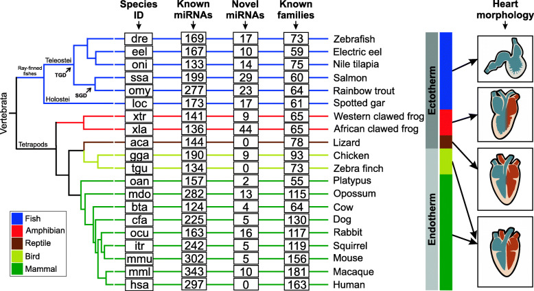
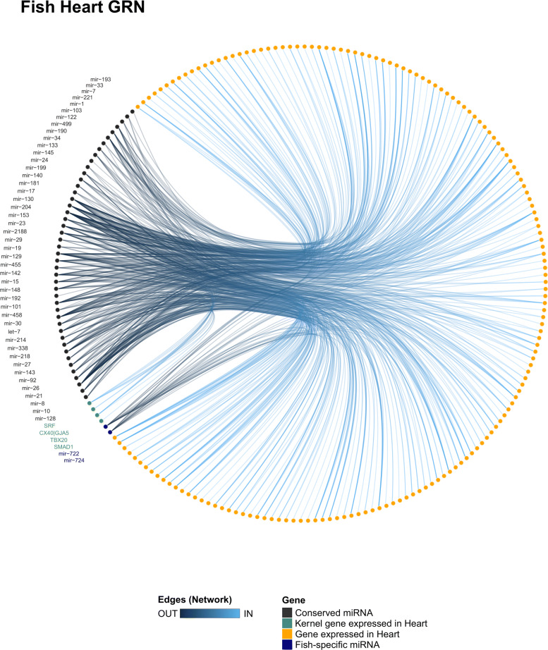
Background: During vertebrate evolution, the heart has undergone remarkable changes that lead to morphophysiological differences in the fully formed heart of these species, such as chamber septation, heart rate frequency, blood pressure, and cardiac output volume. Despite these differences, the heart developmental process is guided by a core gene set conserved across vertebrates. Nonetheless, the regulatory mechanisms controlling the expression of genes involved in heart development and maintenance are largely uncharted. MicroRNAs (miRNAs) have been described as important regulatory elements in several biological processes, including heart biology. These small RNA molecules are broadly conserved in sequence and genomic context in metazoans. Mutations may occur in miRNAs and/or genes that contribute to the establishment of distinct repertoires of miRNA-target interactions, thereby favoring the differential control of gene expression and, consequently, the origin of novel phenotypes. In fact, several studies showed that miRNAs are integrated into genetic regulatory networks (GRNs) governing specific developmental programs and diseases. However, studies integrating miRNAs in vertebrate heart GRNs under an evolutionary perspective are still scarce.

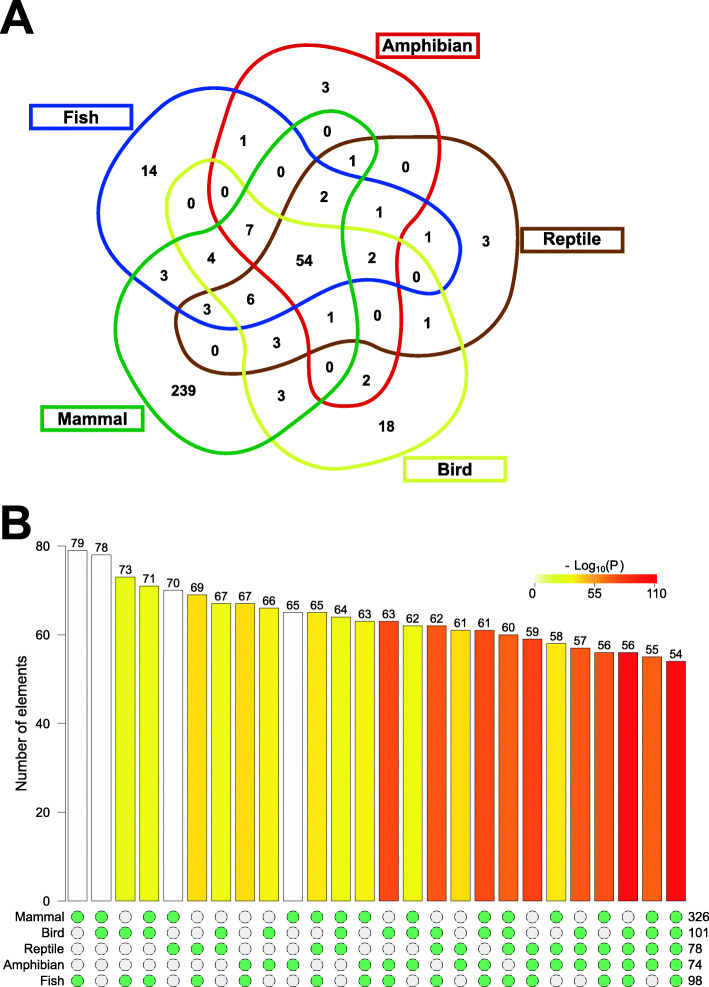
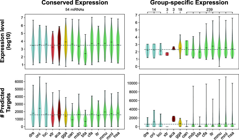
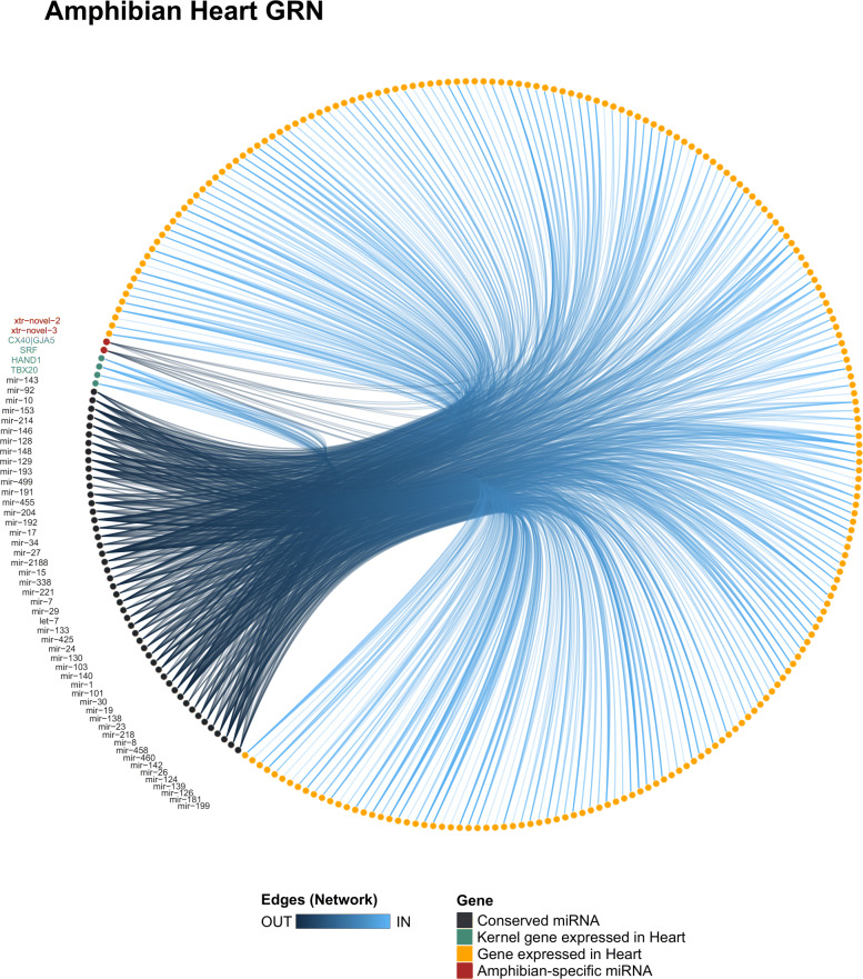
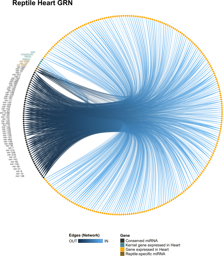
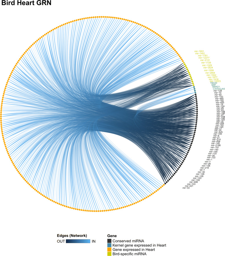
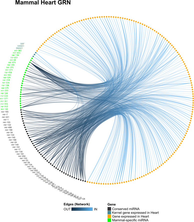
Results: We comprehensively examined and compared the heart miRNome of 20 species representatives of the five major vertebrate groups. We found 54 miRNA families with conserved expression and a variable number of miRNA families with group-specific expression in fishes, amphibians, reptiles, birds, and mammals. We also detected that conserved miRNAs present higher expression levels and a higher number of targets, whereas the group-specific miRNAs present lower expression levels and few targets.

Conclusions: Both the conserved and group-specific miRNAs can be considered modulators orchestrating the core and peripheral genes of heart GRNs of vertebrates, which can be related to the morphophysiological differences and similarities existing in the heart of distinct vertebrate groups. We propose a hypothesis to explain evolutionary differences in the putative functional roles of miRNAs in the heart GRNs analyzed. Furthermore, we present new insights into the molecular mechanisms that could be helping modulate the diversity of morphophysiology in the heart organ of vertebrate species.

Keywords: Cardiac miRNAs; Comparative genomics; Functional genomics; Genetic regulatory network; Non-coding RNA; Small RNA.


Conflict of interest statement

The authors declare that they have no competing interests.

Figures

Fig. 1
Fig. 1
Heart miRNAs in vertebrates. Species ID is indicated at left. Known miRNAs are miRNAs with orthologs identified based on sequence similarity with miRNAs annotated in miRBase and MirGeneDB. Novel miRNAs are putative miRNAs identified in each species by our pipeline. Known families are based on miRBase and MirGeneDB annotations. Heart Morphology is a simplified representation of heart for each group of vertebrates (fishes: two-chambered heart and ectothermy; amphibians: three-chambered heart and ectothermy; reptiles: representative of the Lepidossauria clade presenting a three-chambered heart with partial division at the ventricle and ectothermy; birds: representatives of the Archosauria clade with four-chambered heart and endothermy; mammals: four-chambered heart and endothermy). TGD is Teleost-specific Genome Duplication. SGD is Salmonid-specific Genome Duplication. The phylogenetic tree is a handmade tree derived by merging tree available at TimeTree resource [30] and trees published by [–33]
Fig. 2
Fig. 2
Intersections of vertebrate heart miRNA expression profile. (a) Venn diagram showing the intersections of vertebrate heart miRNA families. (b) Fisher’s exact test results for all intersections (p < 0.005 were considered statistically significant). The numbers at the right bottom indicate the number of miRNA families in the groups indicated at the left bottom. The numbers at the top of the bars indicate the number of miRNA families intersecting between the groups included for the statistical tests as stated by the green points at the bottom
Fig. 3
Fig. 3
Expression level and the number of predicted targets of miRNAs with conserved and group-specific expression in the heart of vertebrates. The number of conserved and group-specific miRNAs analyzed in each species is indicated at the top of the plots. Violin plots of the expression level (top) and the number of predicted targets (bottom)
Fig. 4
Fig. 4
Heart GRN of fishes. The fish heart GRN showing all miR-target interactions detected for the conserved (black) and group-specific miRNAs (blue)
Fig. 5
Fig. 5
Heart GRN of amphibians. The amphibian heart GRN showing all miR-target interactions detected for the conserved (black) and group-specific miRNAs (red)
Fig. 6
Fig. 6
Heart GRN of reptiles. The reptile heart GRN showing all miR-target interactions detected for the conserved (black) and group-specific miRNAs (brown)
Fig. 7
Fig. 7
Heart GRN of birds. The bird heart GRN showing all miR-target interactions detected for the conserved (black) and group-specific miRNAs (yellow)
Fig. 8
Fig. 8
Heart GRN of mammals. The mammal heart GRN showing all miR-target interactions detected for the conserved (black) and group-specific miRNAs (green)

References

    1. Olson EN. Gene regulatory networks in the evolution and development of the heart. Science. 2006;313(5795):1922–7. doi: 10.1126/science.1132292. - DOI - PMC - PubMed
    1. Jensen B, Wang T, Christoffels VM, Moorman AF. Evolution and development of the building plan of the vertebrate heart. Biochim Biophys Acta Mol Cell Res. 2013;1833(4):783–94. doi: 10.1016/j.bbamcr.2012.10.004. - DOI - PubMed
    1. Burggren W, Farrell A, Lillywhite H. Vertebrate cardiovascular systems. Compr Physiol. 2010;:215–308.
    1. Jensen B, Nielsen JM, Axelsson M, Pedersen M, Löfman C, Wang T. How the python heart separates pulmonary and systemic blood pressures and blood flows. J Exp Biol. 2010;213(10):1611–7. doi: 10.1242/jeb.030999. - DOI - PubMed
    1. Lillywhite HB, Zippel KC, Farrell AP. Resting and maximal heart rates in ectothermic vertebrates. Comp Biochem Physiol A Mol Integr Physiol. 1999;124(4):369–82. doi: 10.1016/S1095-6433(99)00129-4. - DOI - PubMed

LinkOut - more resources