

Ivy Sign in Moyamoya Disease: A Comparative Study of the FLAIR Vascular Hyperintensity Sign Against Contrast-Enhanced MRI
- PMID: 33664105
- PMCID: PMC8040985
- DOI: 10.3174/ajnr.A7010
Ivy Sign in Moyamoya Disease: A Comparative Study of the FLAIR Vascular Hyperintensity Sign Against Contrast-Enhanced MRI
Abstract
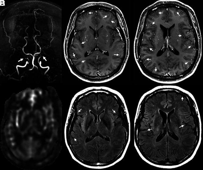
Background and purpose: The ability of the ivy sign on contrast-enhanced T1-weighted MR imaging (CEMR) to reflect cerebral perfusion and postoperative revascularization in Moyamoya disease remains largely unknown. We aimed to compare the capabilities of CEMR and FLAIR.
Materials and methods: CEMR, FLAIR, arterial spin-labeling, and DSA were performed in 44 patients with Moyamoya disease. The ivy sign was scored separately on CEMR and FLAIR using the Alberta Stroke Program Early CT Score. The status of leptomeningeal collaterals was scored on DSA. The postoperative Matsushima grade was evaluated at least 3 months after surgical revascularization.
Results: Scoring of the ivy sign on CEMR showed excellent interrater reliability, and FLAIR vascular hyperintensity showed moderate interrater reliability. Correlation analyses revealed that DSA scores were more consistent with the CEMR-based ivy sign score (r = 0.25, P = .03) than with FLAIR vascular hyperintensity (r = 0.05, P = .65). The CEMR-based ivy sign score was significantly correlated with CBF in late-Suzuki stage Moyamoya disease (t = -2.64, P = .02). The CEMR-based ivy sign score at baseline was significantly correlated with the postoperative Matsushima grade (r = 0.48, P = .03).
Conclusions: In this study, CEMR outperformed FLAIR in capturing the ivy sign in Moyamoya disease. In addition, the CEMR-based ivy sign score provided adequate information on hemodynamic status and postoperative neovascularization. The current study suggested that CEMR could be considered as an alternative to FLAIR in future studies investigating leptomeningeal collaterals in Moyamoya disease.
© 2021 by American Journal of Neuroradiology.
Figures

Comment in
-
Reply.AJNR Am J Neuroradiol. 2021 Sep;42(9):E70. doi: 10.3174/ajnr.A7233. Epub 2021 Jul 15. AJNR Am J Neuroradiol. 2021. PMID: 34266870 Free PMC article. No abstract available.
-
The Possible Difference of Underlying Pathophysiologies between "Ivy Sign" on Contrast-Enhanced MRI and FLAIR.AJNR Am J Neuroradiol. 2021 Sep;42(9):E69. doi: 10.3174/ajnr.A7176. Epub 2021 Jul 15. AJNR Am J Neuroradiol. 2021. PMID: 34266872 Free PMC article. No abstract available.
References
-
- Research Committee on the Pathology and Treatment of Spontaneous Occlusion of the Circle of Willis; Health Labour Sciences Research Grant for Research on Measures for Infractable Diseases. Guidelines for diagnosis and treatment of moyamoya disease (spontaneous occlusion of the circle of Willis). Neurol Med Chir (Tokyo) 2012;52:245–66 10.2176/nmc.52.245 - DOI - PubMed
-
- Kawashima M, Noguchi T, Takase Y, et al. . Unilateral hemispheric proliferation of ivy sign on fluid-attenuated inversion recovery images in moyamoya disease correlates highly with ipsilateral hemispheric decrease of cerebrovascular reserve. AJNR Am J Neuroradiol 2009;30:1709–16 10.3174/ajnr.A1679 - DOI - PMC - PubMed
Publication types
MeSH terms
LinkOut - more resources
Full Text Sources
Other Literature Sources