

The Pharyngolaryngeal Venous Plexus: A Potential Pitfall in Surveillance Imaging of the Neck
- PMID: 33664114
- PMCID: PMC8115379
- DOI: 10.3174/ajnr.A7033
The Pharyngolaryngeal Venous Plexus: A Potential Pitfall in Surveillance Imaging of the Neck
Abstract
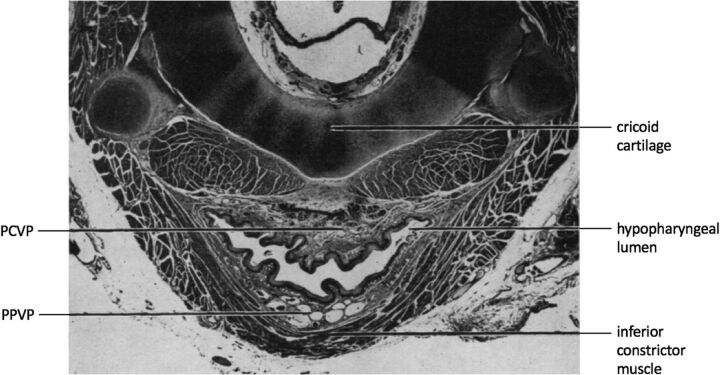
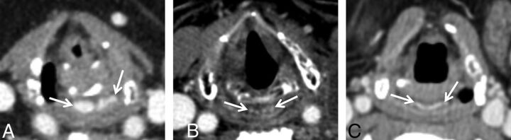
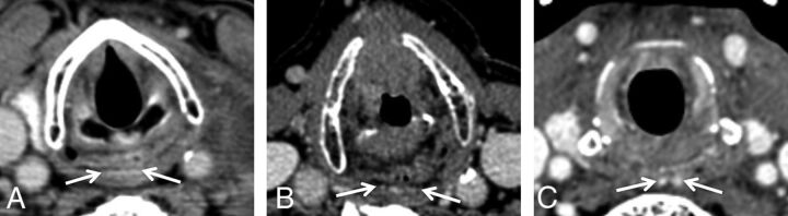
Background and purpose: Among patients undergoing serial neck CTs, we have observed variability in the appearance of the pharyngolaryngeal venous plexus, which comprises the postcricoid and posterior pharyngeal venous plexuses. We hypothesize changes in plexus appearance from therapeutic neck irradiation. The purposes of this study are to describe the CT appearance of the pharyngolaryngeal venous plexus among 2 groups undergoing serial neck CTs-patients with radiation therapy-treated laryngeal cancer and patients with medically treated lymphoma-and to assess for changes in plexus appearance attributable to radiation therapy.
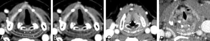
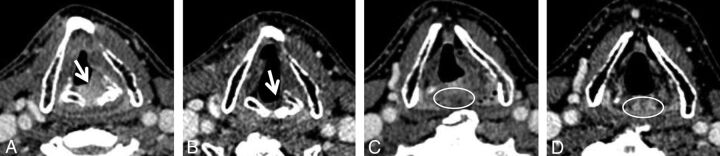
Materials and methods: For this retrospective study of 98 patients (49 in each group), 448 contrast-enhanced neck CTs (222 laryngeal cancer; 226 lymphoma) were assessed. When visible, the plexus anteroposterior diameter was measured, and morphology was categorized.
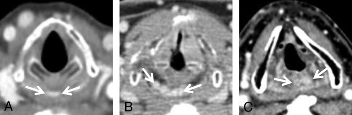
Results: At least 1 plexus component was identified in 36/49 patients with laryngeal cancer and 37/49 patients with lymphoma. There were no statistically significant differences in plexus visibility between the 2 groups. Median anteroposterior diameter was 2.1 mm for the postcricoid venous plexus and 1.6 mm for the posterior pharyngeal venous plexus. The most common morphology was "bilobed" for the postcricoid venous plexus and "linear" for the posterior pharyngeal venous plexus. The pharyngolaryngeal venous plexus and its components were commonly identifiable only on follow-up imaging.
Conclusions: Head and neck radiologists should be familiar with the typical location and variable appearance of the pharyngolaryngeal plexus components so as not to mistake them for neoplasm. Observed variability in plexus appearance is not attributable to radiation therapy.
© 2021 by American Journal of Neuroradiology.
Figures
Similar articles
-
Pharyngolaryngeal Venous Plexus Mimicking Recurrent Hypopharyngeal Cancer.Radiol Imaging Cancer. 2024 Jul;6(4):e240039. doi: 10.1148/rycan.240039. Radiol Imaging Cancer. 2024. PMID: 38940691 Free PMC article. No abstract available.
-
Endoscopic Ho laser interstitial therapy for pharyngolaryngeal venous malformations in adults.Eur Arch Otorhinolaryngol. 2015 Apr;272(4):937-940. doi: 10.1007/s00405-014-3463-y. Epub 2014 Dec 23. Eur Arch Otorhinolaryngol. 2015. PMID: 25534288
-
Pharyngography after head and neck irradiation: differentiation of postirradiation edema from recurrent tumor.AJR Am J Roentgenol. 1993 Dec;161(6):1205-8. doi: 10.2214/ajr.161.6.8249726. AJR Am J Roentgenol. 1993. PMID: 8249726
-
Rare presentations of ordinary lipomas of the head and neck: a review.AJNR Am J Neuroradiol. 1986 Jul-Aug;7(4):657-64. AJNR Am J Neuroradiol. 1986. PMID: 3088944 Free PMC article. Review.
-
Postoperative Pharynx and Larynx.Neuroimaging Clin N Am. 2022 Feb;32(1):37-53. doi: 10.1016/j.nic.2021.08.009. Neuroimaging Clin N Am. 2022. PMID: 34809843 Review.
Cited by
-
New, safe and simple endoscopic cricopharyngeal myotomy with a curved rigid laryngoscope: A case report.Mol Clin Oncol. 2023 Jan 10;18(2):10. doi: 10.3892/mco.2023.2606. eCollection 2023 Feb. Mol Clin Oncol. 2023. PMID: 36761390 Free PMC article.
-
Pharyngolaryngeal Venous Plexus Mimicking Recurrent Hypopharyngeal Cancer.Radiol Imaging Cancer. 2024 Jul;6(4):e240039. doi: 10.1148/rycan.240039. Radiol Imaging Cancer. 2024. PMID: 38940691 Free PMC article. No abstract available.
References
-
- Bourgery J, Jacob N. Atlas of Human Anatomy and Surgery: The Complete Colored Plates of 1831–1854. 25th ed. Taschen; 2005
-
- von Luschka H. Der Kehlkopf des Menschen. H. Laupp; 1871: 147
-
- Bimar L, Lapeyre JM. Recherches sur les veines du pharynx. Comp Rend Acad d Sc 1887;105:825
-
- Elze C. Die venosen Wundernetze der Pars laryngea pharyngis. Anat Anz 1918;51:205–07
MeSH terms
LinkOut - more resources
Full Text Sources
Other Literature Sources
Medical
Research Materials