. 2021 Mar 4;12(3):240.
doi: 10.1038/s41419-021-03524-y.

Mesenchymal stem cells alleviate experimental immune-mediated liver injury via chitinase 3-like protein 1-mediated T cell suppression

Affiliations

Mesenchymal stem cells alleviate experimental immune-mediated liver injury via chitinase 3-like protein 1-mediated T cell suppression

Qiuli Liu et al. Cell Death Dis. .

Abstract

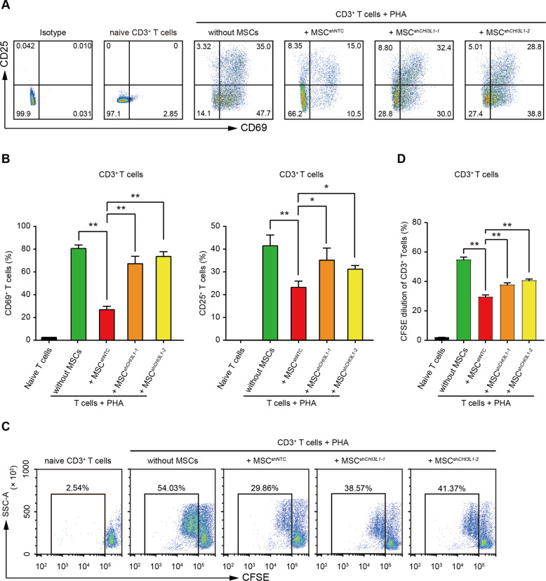
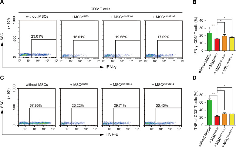
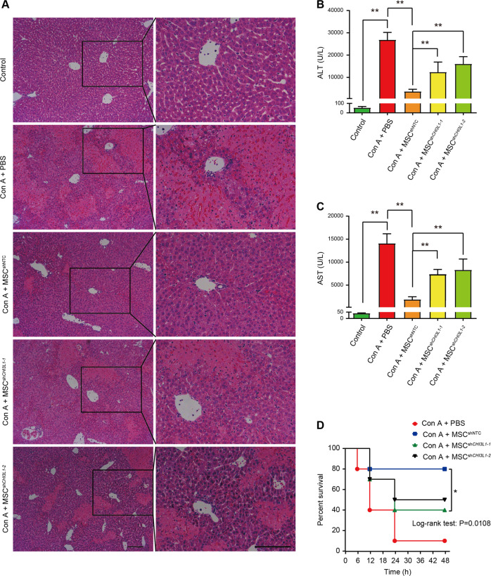
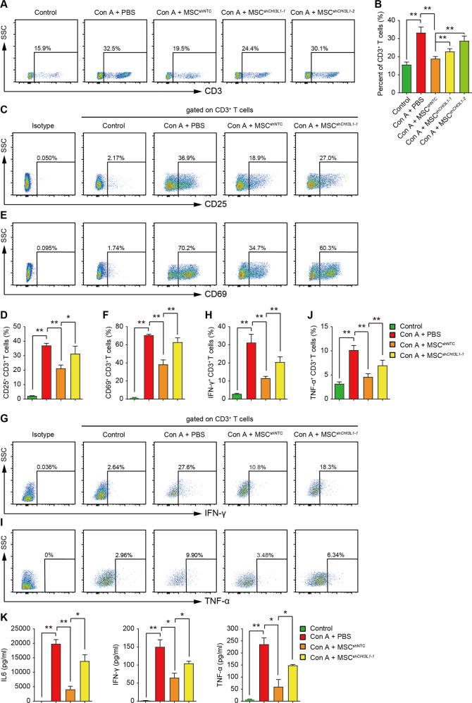
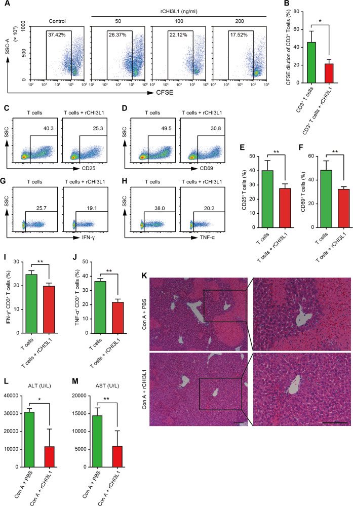
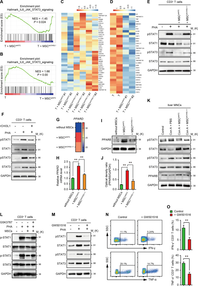
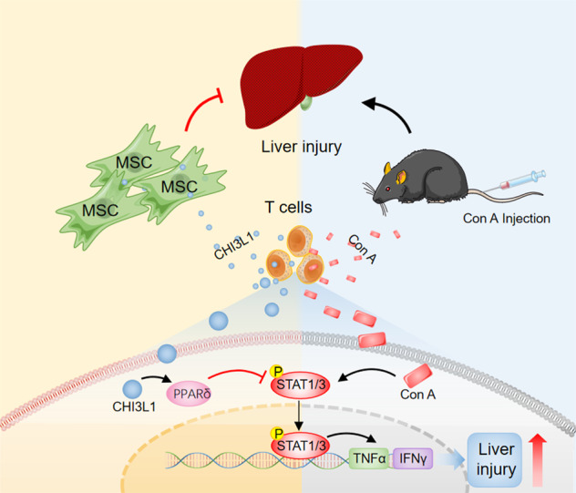
Liver diseases with different pathogenesis share common pathways of immune-mediated injury. Chitinase-3-like protein 1 (CHI3L1) was induced in both acute and chronic liver injuries, and recent studies reported that it possesses an immunosuppressive ability. CHI3L1 was also expressed in mesenchymal stem cells (MSCs), thus we investigates the role of CHI3L1 in MSC-based therapy for immune-mediated liver injury here. We found that CHI3L1 was highly expressed in human umbilical cord MSCs (hUC-MSCs). Downregulating CHI3L1 mitigated the ability of hUC-MSCs to inhibit T cell activation, proliferation and inflammatory cytokine secretion in vitro. Using Concanavalin A (Con A)-induced liver injury mouse model, we found that silencing CHI3L1 significantly abrogated the hUC-MSCs-mediated alleviation of liver injury, accompanying by weakened suppressive effects on infiltration and activation of hepatic T cells, and secretion of pro-inflammatory cytokines. In addition, recombinant CHI3L1 (rCHI3L1) administration inhibited the proliferation and function of activated T cells, and alleviated the Con A-induced liver injury in mice. Mechanistically, gene set enrichment analysis showed that JAK/STAT signalling pathway was one of the most significantly enriched gene pathways in T cells co-cultured with hUC-MSCs with CHI3L1 knockdown, and further study revealed that CHI3L1 secreted by hUC-MSCs inhibited the STAT1/3 signalling in T cells by upregulating peroxisome proliferator-activated receptor δ (PPARδ). Collectively, our data showed that CHI3L1 was a novel MSC-secreted immunosuppressive factor and provided new insights into therapeutic treatment of immune-mediated liver injury.


Conflict of interest statement

The authors declare no competing interests.

Figures

Fig. 1
Fig. 1. hUC-MSCs inhibit the activation and proliferation of T cells through CHI3L1 in vitro.
A Representative flow cytometry plots of CD69 and CD25 expression on CD3+ T cells in indicated groups. CD3+CD25- T cells were sorted by FACS and cultured alone (naïve T cells) or activated with PHA (2.5 μg/mL) for 24 h. For co-culture, sorted T cells were co-cultured with MSCs in the presence of PHA for 24 h. Without MSCs, activated T cells cultured alone; + MSCshNTC, activated T cells co-cultured with hUC-MSCs transduced with shNTC; + MSCshCHI3L1, activated T cells co-cultured with hUC-MSCs transduced with shRNA for CHI3L1. B Quantification of CD69 (left) and CD25 (right) positive CD3+ T cells in (A). C Representative flow cytometry plots and quantification (D) of flow cytometry analysis for proliferation of CD3+ T cells labelled with CFSE in indicated groups. Data in (B) and (D) are shown as mean ± SD (n = 3 biological replicates) with the indicated significance (*p < 0.05, **p < 0.01).
Fig. 2
Fig. 2. hUC-MSCs downregulate the pro-inflammatory cytokine production of CD3+ T cells partially through CHI3L1 in vitro.
A Representative plots and quantification (B) of flow cytometry analysis for IFN-γ-producing CD3+ T cells in indicated groups. C Representative plots and quantification (D) of flow cytometry analysis for TNF-α-producing CD3+ T cells in indicated groups. Flow cytometry analysis were conducted 3 days after CD3+ T cells co-cultured with MSCs. Data in (B) and (D) are shown as mean ± SD (n = 3 biological replicates) with the indicated significance (*p < 0.05, **p < 0.01).
Fig. 3
Fig. 3. hUC-MSCs alleviate Con A-induced mouse liver injury through CHI3L1.
A Representative Hematoxylin and eosin (H&E) staining photographs of liver tissues 24 h after Con A administration in indicated groups. Scale bar = 100 μm. B Serum ALT and AST (C) levels were measured 24 h after Con A treatment in indicated groups. D The survival curves of mice in indicated groups (data were collected every 6 h, n = 10 for each group). Data in (B) and (C) are shown as mean ± SD (n = 4–7 for each group) with indicated significance (*p < 0.05, **p < 0.01).
Fig. 4
Fig. 4. hUC-MSCs suppress the activation and pro-inflammatory cytokine secretion of hepatic T cells through CHI3L1 in Con A-induced hepatitis.
A Representative plots and quantification (B) of flow cytometry analysis for CD3+ T cells in liver MNCs 24 h after Con A administration in indicated groups. CF Representative plots and quantification of flow cytometry analysis for CD25 (CD) and CD69 (EF) expression in CD3+ T cells in liver MNCs 24 h after Con A administration in indicated groups. GJ Representative plots and quantification of flow cytometry analysis for IFN-γ (GH) and TNF-α (IJ) -producing CD3+ T cells in liver MNCs 24 h after Con A administration in indicated groups. K Serum concentrations of IL6, IFN-γ, and TNF-α 24 h after Con A administration in indicated groups were detected by CBA kit. Analyses in (GJ) were conducted with liver MNCs stimulated with PMA (50 ng/mL), ionomycin (500 ng/mL) and BFA (10 μg/mL) for 6 h after isolation. Data in (B), (D), (F), (H), (J), and (K) are shown as mean ± SD (n = 5 for each group) with indicated significance (*p < 0.05, **p < 0.01).
Fig. 5
Fig. 5. Recombinant CHI3L1 (rCHI3L1) ameliorates Con A-induced hepatitis.
A Representative flow cytometry plots for analysing proliferation of CD3+ T cells labelled with CFSE treated with rCHI3L1 in indicated conditions. PBS was used as a control. B Quantification of flow cytometry analysis for proliferation of CD3+ T cells labelled with CFSE in PBS (control) or rCHI3L1 (100 ng/mL) treated group (n = 3 biological replicates). CF Representative plots (CD) and quantification (EF) of flow cytometry analysis for CD25 and CD69 expression in CD3+ T cells treated with control (PBS) or rCHI3L1 (100 ng/mL) (n = 3 biological replicates). GJ Representative plots (GH) and quantification (IJ) of flow cytometry analysis for IFN-γ and TNF-α expression in CD3+ T cells treated with control (PBS) or rCHI3L1 (100 ng/mL) (n = 3 biological replicates). K Representative H&E staining photographs of liver tissues 24 h after Con A administration in control (PBS) and rCHI3L1 (500 ng) treated groups. Scale bar = 100 μm. L Serum ALT and AST (M) levels 24 h after Con A treatment in indicated groups (n = 4–7 mice per group). Data in (B), (E), (F), (I), (J), (L), and (M) are shown as mean ± SD with indicated significance (*p < 0.05, **p < 0.01).
Fig. 6
Fig. 6. hUC-MSC-derived CHI3L1 inhibits T cells through PPARδ/STAT1/3.
A GSEA showing that the IL6-JAK-STAT3 pathway is enriched in T cells co-cultured with MSCshCHI3L1 compared with T + MSCshNTC group. B GSEA showing that the IL6-JAK-STAT3 pathway is repressed in T cells co-cultured with MSCshNTC compared to activated T cells. CD Heatmap of RNA-sequencing data showing the expression level of genes involved in IL6-JAK-STAT3 pathway in indicated samples. E Western blot for phosphorylated and total STAT1 and STAT3 in T cells with indicated treatments for 2 days. F Western blot for phosphorylated and total STAT1 and STAT3 in T cells cultured with or without rCHI3L1 (100 ng/mL). G Heatmap of RNA-sequencing data showing the expression level of PPARD in activated T cells (T cells), T cells co-cultured with MSCshNTC (+ MSCshNTC), and T cells co-cultured with MSCshCHI3L1 (+ MSC shCHI3L1). H RT-qPCR for PPARD in T cells cultured alone or co-cultured with hUC-MSCs with or without CHI3L1 knockdown. I Western blot and corresponding densitometry analysis (J) for PPARδ in T cells culture alone, or co-cultured with MSCshNTC or MSCshCHI3L1. K Western blot for phosphorylated and total STAT1 and STAT3 in liver MNCs in indicated groups. L Representative western blot for testing phosphorylated-STAT1 (p-STAT1), total STAT1, phosphorylated-STAT3 (p-STAT3), and total STAT3 in human naïve CD3+ T cells, CD3+ T cells activated by PHA, activated CD3+ T cells co-cultured with MSCs, and activated CD3+ T cells co-cultured with MSCs in the presence of PPARδ inhibitor (GSK3787, S8025, Selleck). GSK3787 was added simultaneously with PHA at a concentration of 1 μM for 48 h. M Western blot for phosphorylated and total STAT1 and STAT3 in T cells stimulated with PHA in the absence or presence of PPARδ agonist GW501516 (10 μM). N Representative flow cytometry plots and quantification (O) of IFN-γ (upper) and TNF-α (lower)-producing CD3+ T cells treated with GW501516. DMSO was used as a control in (LO).
Fig. 7
Fig. 7. hUC-MSCs alleviate experimental immune-mediated liver injury via CHI3L1-mediated T cell suppression.
Schematic demonstration of the main message of our work.

Similar articles

Cited by

References

    1. Eksteen B, Afford SC, Wigmore SJ, Holt AP, Adams DH. Immune-mediated liver injury. Semin. Liver Dis. 2007;27:351–366. doi: 10.1055/s-2007-991512. - DOI - PubMed
    1. Wang M, Yuan Q, Xie L. Mesenchymal Stem Cell-Based Immunomodulation: Properties and Clinical Application. Stem Cells Int. 2018;2018:3057624. - PMC - PubMed
    1. Hu C, Zhao L, Li L. Current understanding of adipose-derived mesenchymal stem cell-based therapies in liver diseases. Stem Cell Res Ther. 2019;10:199. doi: 10.1186/s13287-019-1310-1. - DOI - PMC - PubMed
    1. Kholodenko IV, Kurbatov LK, Kholodenko RV, Manukyan GV, Yarygin KN. Mesenchymal Stem Cells in the Adult Human Liver: Hype or Hope? Cells. 2019;8:1127. doi: 10.3390/cells8101127. - DOI - PMC - PubMed
    1. de Miguel MP, Prieto I, Moratilla A, Arias J, Aller MA. Mesenchymal Stem Cells for Liver Regeneration in Liver Failure: From Experimental Models to Clinical Trials. Stem Cells Int. 2019;2019:3945672. doi: 10.1155/2019/3945672. - DOI - PMC - PubMed

Publication types

MeSH terms