. 2021 Mar 4;12(3):241.
doi: 10.1038/s41419-021-03526-w.

Impairment in inflammasome signaling by the chronic Pseudomonas aeruginosa isolates from cystic fibrosis patients results in an increase in inflammatory response

Affiliations

Impairment in inflammasome signaling by the chronic Pseudomonas aeruginosa isolates from cystic fibrosis patients results in an increase in inflammatory response

Melissa S Phuong et al. Cell Death Dis. .

Abstract

Pseudomonas aeruginosa is a common respiratory pathogen in cystic fibrosis (CF) patients which undergoes adaptations during chronic infection towards reduced virulence, which can facilitate bacterial evasion of killing by host cells. However, inflammatory cytokines are often found to be elevated in CF patients, and it is unknown how chronic P. aeruginosa infection can be paradoxically associated with both diminished virulence in vitro and increased inflammation and disease progression. Thus, we investigated the relationship between the stimulation of inflammatory cell death pathways by CF P. aeruginosa respiratory isolates and the expression of key inflammatory cytokines. We show that early respiratory isolates of P. aeruginosa from CF patients potently induce inflammasome signaling, cell death, and expression of IL-1β by macrophages, yet little expression of other inflammatory cytokines (TNF, IL-6 and IL-8). In contrast, chronic P. aeruginosa isolates induce relatively poor macrophage inflammasome signaling, cell death, and IL-1β expression but paradoxically excessive production of TNF, IL-6 and IL-8 compared to early P. aeruginosa isolates. Using various mutants of P. aeruginosa, we show that the premature cell death of macrophages caused by virulent bacteria compromises their ability to express cytokines. Contrary to the belief that chronic P. aeruginosa isolates are less pathogenic, we reveal that infections with chronic P. aeruginosa isolates result in increased cytokine induction due to their failure to induce immune cell death, which results in a relatively intense inflammation compared with early isolates.


Conflict of interest statement

The authors declare no competing interests.

Figures
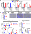
Fig. 1
Fig. 1. P. aeruginosa isolates from chronic CF infections induce less cell death but increased expression of TNF, IL-8, and IL-6 compared with early isolates.
Early isolates were defined as isolates from the first positive P. aeruginosa-positive sputum culture, while chronic isolates were from patients who had 4 years of positive sputum cultures. Primary human macrophages were seeded at 50,000 cells/well and infected with the indicated early and chronic isolates of P. aeruginosa from early and chronic infections or PA14 (10 MOI). Cell viability was measured via neutral red assay and cytokine production was measured via ELISA at 3 h postinfection (A). THP-1 macrophages were seeded in fresh media at 100,000 cells/well in a 96-well plate and infected with P. aeruginosa isolates from CF patients with early or chronic respiratory infections at 1 MOI as described in Methods and in experimental triplicates. At 3 h postinfection, cell viability was evaluated by neutral red assay, and cytokine production was measured in the supernatants by ELISA (B, D). Student’s t-tests were used to compare the means for cell viability, TNF, and IL-8 while Mann–Whitney U tests were used for IL-1β, IL-6, and IL-10. THP-1 macrophages were seeded at 500,000 cells/well and infected with P. aeruginosa (early or late isolate) at 10 MOI. At 2 h postinfection, cells were stained with Hoechst/PI to distinguish live (blue) from dead (pink) cells (C). NuLi-1 bronchial epithelial cells were seeded at 30,000 cells/well in a collagen-coated 96-well plate and infected with the same cohort of early and chronic P. aeruginosa isolates at 100 MOI and in experimental triplicates. Cell viability via neutral red assay and cytokine production in supernatants via ELISA were measured 6 h postinfection (E). Mann–Whitney U tests were used to compare IL-1β and IL-6 while Welch’s t-test was used to compare the means for cell viability. Mean ± SD of triplicates of a representative, or a pool of >3 experiments are shown. (*P < 0.05, ***P < 0.001, ****P < 0.0001).
Fig. 2
Fig. 2. Cell death induced by P. aeruginosa, but not IL-1β expression, is independent of caspase-1.
THP-1 macrophages were seeded at 500,000 cells/well in a 24-well plate. At 1 h postinfection (1 MOI) with the indicated acute and chronic isolates of P. aeruginosa from CF patients. RNA was extracted and cDNA was synthesized. qPCR was conducted on samples targeting genes for various cytokines, and mRNA levels were normalized to β-actin (AD). THP-1 macrophages were seeded at 500,000 cells/well in a 24-well plate. At 3 h postinfection (1 MOI) with the indicated acute and chronic isolates of P. aeruginosa from CF patients, western blotting was performed on cell supernatants or extracts using specific antibodies shown in figure (E). Densitometric analysis of western blots shown in panel E was conducted (F). THP-1 WT and THP1-defCASP1 were infected with PA14 or PAO1 (1 MOI) and western blotting was performed on supernatants or cell extracts at 3 h postinfection (G). THP-1 WT and THP1-defCASP1 were infected with early and chronic P. aeruginosa CF isolates (1 MOI) in experimental triplicates. Cell viability was evaluated at 3 h postinfection by neutral red assay, and IL-1β production was measured in supernatants by ELISA (H). Mann–Whitney U tests were conducted. THP-1 WT and THP-1-defCASP1 macrophages were infected with PA14 or PAO1 P. aeruginosa, and cell viability and cytokine production were measured as mentioned above (I, J). Mean values were compared by Student’s t-tests, except for measured IL-1β for PA14 at 1 MOI and PAO1 at 0.1 MOI, where Welch’s t-tests were used. Mean ± SD of triplicates of a representative, or a pool of >3 experiments are shown. (*P < 0.05, **P < 0.01, ***P < 0.001, ****P < 0.0001).
Fig. 3
Fig. 3. Early CF infection P. aeruginosa isolates and PAO1 are partially dependent on NLRP3 to induce cell death and IL-1β production.
THP-1 macrophages were seeded as described in Fig. 1. THP-1 macrophages were treated with 10 µM of MCC950 at the time of infection with early P. aeruginosa isolates from CF patients and the indicated laboratory reference strains to inhibit NLRP3 activation when infecting THP-1 macrophages. At 3 h postinfection, cell viability was evaluated by neutral red assay (A, G, I) and IL-1β production was measured in cell supernatants by ELISA (B, H, J). THP-1 macrophages were seeded as described in Fig. 1 and pre-treated with 5 µM GSK872 (RipK3 inhibitor), 10 µM z-IETD-FMK (caspase-8 inhibitor) or a DMSO control 2 h prior to infection. Respective treatments were continued during THP-1 macrophage infections with indicated early P. aeruginosa isolates or reference strains (1 MOI). At 3 h postinfection, cell viability was evaluated by neutral red assay (C, E) and IL-1β production was measured in cell supernatants by ELISA (D, F). Mean ± SD of experimental triplicates are shown. Student’s t-tests (A, B, GJ) and one-way ANOVAs followed by Dunnett’s multiple comparisons tests (D, F) were conducted. Mean ± SD of triplicates of a representative experiment are shown. (*P < 0.05, **P < 0.01, ***P < 0.001, ****P < 0.0001).
Fig. 4
Fig. 4. Chronic CF infection P. aeruginosa isolates induce increased activation of NF-κB and MAPK more than the early infection isolates.
THP-1 macrophages were seeded at 500,000 cells/well in a 24-well plate. Immunoblotting was conducted on lysates collected at 1 h postinfection with the indicated CF P. aeruginosa isolates at 1 MOI (A). Densitometric analysis of western blots shown in panel A was performed (B, D, E). THP-1 Lucia™ NF-κB cells were differentiated and incubated with 1 MOI of heat-killed early or chronic CF P. aeruginosa isolates in experimental triplicates. At 3 h, supernatant luciferase activity (relative light units, RLUs) was measured to assess NF-κB activation (C). THP-1 macrophages were seeded at 100,000 cells/well in a 96-well plate. Ralimetinib, a p38 MAPK inhibitor, was added during infections with select chronic P. aeruginosa isolates from CF patients to a final concentration of 0.1 µM. Cell viability via neutral red assay (F) and cytokine production via ELISA (GI) were then measured 3 h postinfection. Mean ± SD of triplicates of a representative experiment are shown. Mean values were compared by Student’s t-tests. (*P < 0.05, **P < 0.01, ***P < 0.001, ****P < 0.0001).
Fig. 5
Fig. 5. Environmental and clinical, non-CF P. aeruginosa isolates also generally exhibit an inverse relationship between induction of cell death and TNF and IL-8 expression.
THP-1 macrophages were seeded as described in Fig. 1. Environmental P. aeruginosa isolates from river water (A) and clinical P. aeruginosa isolates from non-CF patients (i.e., blood, wound, and urinary tract infections) (B) were used to infect THP-1 macrophages at 1 MOI. Cell viability and IL-1β, TNF, and IL-8 production were measured at 3 h postinfection as described in Fig. 1. Simple linear regression analyses were conducted to determine the relationship between cell viability and cytokine expression for infections with (C) environmental and (D) non-CF isolates. Mean ± SD of triplicates of a pool of >3 experiments are shown.
Fig. 6
Fig. 6. PA14 mutants with impaired T3SS induce less cell death but higher expression of TNF and IL-8 compared to wild-type PA14.
Primary human macrophages were seeded at 50,000 cells/well and infected with PA14 or PAO1 at MOI 10. Cell viability via neutral red assay and cytokine production via ELISA were measured 3 h postinfection (A). THP-1 macrophages were seeded as described in Fig. 1. In vitro infections were conducted with wild-type PA14 and PAO1, and the various transposon mutants (fliC::tn, popB::tn, popD::tn, and exsA::tn) of PA14. Cell viability was measured by neutral red assay and cytokine production was evaluated by ELISA at 3 h (B, D) or at 1-h intervals (MOI 10) (C, E). Mean ± SD of triplicates of a representative, or a pool of >3 experiments are shown. One-way ANOVAs were conducted to compare mean values at MOIs of 1 and 10. (****P < 0.0001).
Fig. 7
Fig. 7. CFTR inhibition in THP-1 macrophages does not impact cell death or cytokine production.
THP-1 macrophages were seeded as described in Fig. 1 with the addition of either a DMSO vehicle control or 10 µM CFTR(inh)-172 overnight. In vitro infections were conducted with PA14 (AD) or PAO1 (EH) with the addition of either treatment. At 3 h postinfection, cell viability was evaluated by neutral red assay (A, E) and cytokine production was measured by ELISA (BD, FH). Mean ± SD of triplicates of a representative experiment are shown.
Fig. 8
Fig. 8. PA14 and S. aureus demonstrate an inverse relationship between cell death and cytokine expression.
THP-1 macrophages were seeded as described in Fig. 1 and infected with either PA14 or S. aureus 6538 at various MOIs. Three hours postinfection, cell viability via neutral red and TNF and IL-1β in the supernatant via ELISAs were measured (A). Infections were also performed using an MOI of 1, and cell viability and cytokines were measured at 1-h intervals for up to 3 h (B). Mean ± SD of triplicates of a representative experiment are shown.

Similar articles

Cited by

References

    1. Hartl D, et al. Innate immunity in cystic fibrosis lung disease. J. Cyst. Fibros. 2012;11:363–382. doi: 10.1016/j.jcf.2012.07.003. - DOI - PubMed
    1. Fink SL, Cookson BT. Apoptosis, pyroptosis, and necrosis: mechanistic description of dead and dying eukaryotic cells. Infect. Immun. 2005;73:1907–1916. doi: 10.1128/IAI.73.4.1907-1916.2005. - DOI - PMC - PubMed
    1. Balakrishnan A, Karki R, Berwin B, Yamamoto M, Kanneganti T-D. Guanylate binding proteins facilitate caspase-11-dependent pyroptosis in response to type 3 secretion system-negative Pseudomonas aeruginosa. Cell Death Discov. 2018;4:66–66. doi: 10.1038/s41420-018-0068-z. - DOI - PMC - PubMed
    1. Faure E, Kwong K, Nguyen D. Pseudomonas aeruginosa in chronic lung infections: how to adapt within the host? Front. Immunol. 2018;9:2416. doi: 10.3389/fimmu.2018.02416. - DOI - PMC - PubMed
    1. Amiel E, Lovewell RR, O’Toole GA, Hogan DA, Berwin B. Pseudomonas aeruginosa evasion of phagocytosis is mediated by loss of swimming motility and is independent of flagellum expression. Infect. Immun. 2010;78:2937–2945. doi: 10.1128/IAI.00144-10. - DOI - PMC - PubMed

Publication types

MeSH terms