. 2021 Mar 4;12(3):239.
doi: 10.1038/s41419-021-03521-1.

E3 ubiquitin ligase RNF126 affects bladder cancer progression through regulation of PTEN stability

Affiliations

E3 ubiquitin ligase RNF126 affects bladder cancer progression through regulation of PTEN stability

Huimin Xu et al. Cell Death Dis. .

Abstract

E3 ubiquitin ligase RNF126 (ring finger protein 126) is highly expressed in various cancers and strongly associated with tumorigenesis. However, its specific function in bladder cancer (BCa) is still debatable. Here, we found that RNF126 was significantly upregulated in BCa tissue by TCGA database, and our studies indicated that downregulation of RNF126 significantly inhibited cell proliferation and metastasis through the EGFR/PI3K/AKT signaling pathway in BCa cells. Furthermore, we identified PTEN, an inhibitor of the PI3K/AKT signaling pathway, as a novel substrate for RNF126. By co-immunoprecipitation assays, we proved that RNF126 directly interacts with PTEN. Predominantly, PTEN binds to the C-terminal containing the RING domain of RNF126. The in vivo ubiquitination assay showed that RNF126 specifically regulates PTEN stability through poly-ubiquitination. Furthermore, PTEN knockdown restored cell proliferation, metastasis, and tumor formation of BCa cells inhibited by RNF126 silencing in vitro and in vivo. In conclusion, these results identified RNF126 as an oncogene that functions through ubiquitination and degradation of PTEN in BCa.


Conflict of interest statement

The authors declare no competing interests.

Figures
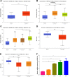
Fig. 1
Fig. 1. Expression of RNF126 in BCa of TCGA samples and different cell lines.
A RNF126 expression in the normal and primary tumor of the bladder. B RNF126 expression is based on different histological subtypes: papillary tumor and non-papillary tumor. C Expression of RNF126 in BCa based on the patient’s race. D Promoter methylation level of RNF126 in BCa. E RNF126 promoter methylation profile is based on histological subtypes. F The qRT-PCR analysis confirmed the transcription level of RNF126 in normal bladder epithelial cells SV-HUC-1 and various BCa cell lines (T24, UMUC3, 5637, BIU78). ***p < 0.001, **p < 0.01, *p < 0.05.
Fig. 2
Fig. 2. Depletion of RNF126 inhibited BCa cell proliferation, migration, and viability.
A The qRT-PCR verified the efficiency of knockdown RNF126 by siRNA (RNF126-si1 and RNF126-si2) in two BCa cells T24 and UMUC3. B The western blotting verified the expression of RNF126 downregulation in T24 and UMUC3 cells after transfection with the RNF126 siRNA. The internal control was GAPDH abundance. C, D The MTT assay evaluated the T24 and UMUC3 cells growth and viability from day 1 until day 5 after transfection with the RNF126-si1 and si2 (orange line and green line). E The colony formation assay showed the effect of RNF126 knockdown on the cell survival of UMUC3 and T24 cells after the transfection. Scale bar = 1 cm. F Colony numbers were counted and plotted as indicated. G The transwell assay evaluated cell migration of the RNF126-si1, si2 and NC treated BCa cells. Scale bar = 100 µm. H The number of cell migration was counted and statistically analyzed. I The wound healing assay evaluated the migration of siRNF126-treated BCa cells after scratching for 24 h. Scale bar = 100 µm. J The western blotting revealed the downregulation of protein abundance of EMT markers (E-Cad, N-Cad) in the BCa by reduction of RNF126. GAPDH severed as control. ***p < 0.001, **p < 0.01, *p < 0.05.
Fig. 3
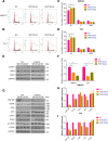
Fig. 3. RNF126 deficiency affects the cell cycle and EGFR/PI3K/AKT signaling pathway in BCa cells.
A, B The flow cytometry analysis demonstrated the percentage (%) of cells in different phases of cell cycle. The T24 and UMUC3 cells were treated with RNF126-si1 and si2 for two days. C, D The percentage (%) of cells in each phase was statistically analyzed from three independent experiments. E The western blotting revealed downregulation of protein associated with the cell cycle (CCND1, CDK2 and CDK4) in the BCa cells after silencing RNF126. The internal control was GAPDH. F The relative mRNA level of CCND1 in different cell types, treatment of siRNA. H The western blotting showed the protein level of EGFR, PI3K, total and phosphorylated AKT, mTOR, and PTEN in BCa cells treated with RNF126-si1and si2. The internal control was GAPDH. I The relative mRNA level of EGFR, PI3K, AKT, PTEN in different cell types, and siRNA treatment. ***p < 0.001, **p < 0.01, *p < 0.05.
Fig. 4
Fig. 4. RNF126 interacts with PTEN.
A The co-immunoprecipitation of GFP-PTEN and FLAG-RNF126 in 293T cells. The 293T cells were transfected with FLAG-RNF126 and GFP-PTEN plasmids for 36 h. B The endogenous co-immunoprecipitation of PTEN and RNF126 in T24 cells. C–E The interactions between full-length RNF126 and truncated forms of GFP-PTEN were observed by co-immunoprecipitation in 293T cells. F The interactions between full-length PTEN and truncated forms of FLAG-RNF126 were observed by co-immunoprecipitation in 293T cells. G Protein domains of RNF126 and PTEN. Numbers indicate how long the proteins were cut off. RNF126 protein was truncated between the RING finger domain and the Zn finger domain. PTEN protein was truncated to reserve the C2 domain.
Fig. 5
Fig. 5. RNF126 regulates the stability of PTEN.
A The 293T cells were transfected with GFP-PTEN and FLAG-RNF126 plasmids at different doses 36 h, then the cells were collected, and anti-GFP antibodies were detected by western blotting to determine the level of exogenous PTEN. B GFP-PTEN and FLAG-RNF126 were transfected into 293T cells for 24 h, then the cells were treated with dimethyl sulphoxide (DMSO), 10 μM MG132 (#S2619, Selleck) or 20 μM chloroquine (CQ, #S8808, Selleck) for 8 h. The cells were collected and anti-GFP antibodies were detected by western blot. C FLAG-RNF126 plasmid was transfected into 293T cells, and then 100 μg/ml cycloheximide (CHX, #S7418, Selleck) was respectively added at the specified time points for 0 h, 4 h, 8 h, and 12 h. Then, the cells were harvested, and western blot detected GFP antibodies to determine the half-life of GFP-PTEN protein. D The RNF126 siRNA was transfected into BCa 5637 cells for 36 h, and then 100 μg/ml CHX was added at specified time points for 0 h, 4 h, 8 h, 12 h, and collect cells. The half-life of endogenous PTEN protein was determined by western blot. E, F The ImageJ v1.45 software was used to quantified PTEN protein abundance. The relative level of PTEN protein plotted as indicated. G The in vivo ubiquitination assay showed that RNF126 poly-ubiquitinated PTEN. HA-Ub, GFP-PTEN and FLAG-RNF126 plasmids were transfected into 293T cells for 36 h. The cells were treated with 10 μM MG132 or DMSO for 8 h before harvest. H Graphic model of RNF126 affecting proliferation and metastasis through ubiquitination and degradation of PTEN in BCa.
Fig. 6
Fig. 6. Depletion of RNF126 suppressed BCa cell proliferation and metastasis through upregulating PTEN.
A The MTT assay evaluated the growth and viability of the T24 cell from day 1 until day 5. A lentiviral shRNF126 and a lentiviral control group shNC stably transfected T24 cell were established. The cells were transfected with siNC (red line: shNC and orange line: shRNF126) or siPTEN (olive line: shNC + siPTEN and green line: shRNF126 + siPTEN). B, C The flow cytometry analysis demonstrated the distribution of cells in different phases of cell cycle. The four groups were the same as above. D The colony formation assay showed the effect of RNF126 knockdown on the cell survival of UMUC3 and T24 cells after the indicated transfection. Scale bar = 1 cm. E Colony numbers were counted and plotted as indicated. F The transwell assay evaluated cell migration of the RNF126 or PTEN knockdown treated T24 cells. Scale bar = 100 µm. G The relative number of cell migration was statistically analyzed. H The western blotting indicated the protein level of RNF126, PTEN, PI3K, p-AKT, AKT, CCND1, p21, and N-Cad. Loading control was GAPDH. ***p < 0.001, **p < 0.01, *p < 0.05.
Fig. 7
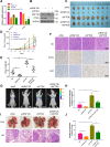
Fig. 7. Effects of RNF126 and PTEN on BCa cell progression in vivo.
A, B The qRT-PCR and western blotting showed the stable knockdown of RNF126 and PTEN mRNA and protein levels in T24 cells. C LV-control, LV-shRNF126, LV-shPTEN and LV-shRNF126 + shPTEN T24 cells were subcutaneously injected into four groups mice to generate the xenograft models. When the mice grew for 5 weeks, the xenografts were taken out and photographed (n = 8). D Tumor volume growth of BCa cells was measured twice a week until it grew for 5 weeks. E After the mice were sacrificed, the tumor weight was measured. F IHC demonstrated the expression of RNF126, PTEN and Ki67 proteins in tumors. Scale bar = 100 μm. G, H The mice’s tails were intravenous injected the LV-control, LV-shRNF126, LV-shPTEN and LV-shRNF126 + shPTEN T24 cells to establish the pulmonary metastasis models (n = 3). The BCa cell migration capacity was demonstrated by the fluorescence of pulmonary metastases. I After injection for 8 weeks, the mice were sacrificed and the lungs were taken out. Then, the lungs were stained with HE. The arrows denote the tumors transferred to the lungs. Scale bar = 100 μm. J The quantification of pulmonary metastases was determined by counting the metastatic nodules. ***p < 0.001, **p < 0.01, *p < 0.05.

Similar articles

Cited by

References

    1. Siegel RL, Miller KD, Jemal A. Cancer statistics, 2020. CA Cancer J. Clin. 2020;70:7–30. doi: 10.3322/caac.21590. - DOI - PubMed
    1. Bray F, et al. Global cancer statistics 2018: GLOBOCAN estimates of incidence and mortality worldwide for 36 cancers in 185 countries. CA Cancer J. Clin. 2018;68:394–424. doi: 10.3322/caac.21492. - DOI - PubMed
    1. Smith AB, et al. Muscle-invasive bladder cancer: evaluating treatment and survival in the National Cancer Data Base. BJU Int. 2014;114:719–726. doi: 10.1111/bju.12601. - DOI - PubMed
    1. Burger M, et al. Epidemiology and risk factors of urothelial bladder cancer. Eur. Urol. 2013;63:234–241. doi: 10.1016/j.eururo.2012.07.033. - DOI - PubMed
    1. Soloway MS. Bladder cancer: lack of progress in bladder cancer–what are the obstacles? Nat. Rev. Urol. 2013;10:5–6. doi: 10.1038/nrurol.2012.219. - DOI - PubMed

Publication types

MeSH terms