. 2021 Mar 4;12(3):245.
doi: 10.1038/s41419-021-03523-z.

Long noncoding RNA LINC00518 induces radioresistance by regulating glycolysis through an miR-33a-3p/HIF-1α negative feedback loop in melanoma

Affiliations

Long noncoding RNA LINC00518 induces radioresistance by regulating glycolysis through an miR-33a-3p/HIF-1α negative feedback loop in melanoma

Yan Liu et al. Cell Death Dis. .

Abstract

The long noncoding RNA, LINC00518, is highly expressed in various types of cancers and is involved in cancer progression. Although LINC00518 promotes the metastasis of cutaneous malignant melanoma (CMM), the mechanism underlaying its effects on CMM radiosensitivity remains unclear. In this study, LINC00518 expression was significantly upregulated in CMM samples, and LINC00518 levels were associated with poor prognosis of patients with CMM. Knockdown of LINC00518 in CMM cells significantly inhibited cell invasion, migration, proliferation, and clonogenicity. LINC00518-mediated invasion, migration, proliferation, and clonogenicity were negatively regulated by the microRNA, miR-33a-3p, in vitro, which increased sensitivity to radiotherapy via inhibition of the hypoxia-inducible factor 1α (HIF-1α)/lactate dehydrogenase A glycolysis axis. Additionally, HIF-1α recognized the miR-33a-3p promoter region and recruited histone deacetylase 2, which decreased the expression of miR-33a-3p and formed an LINC00518/miR-33a-3p/HIF-1α negative feedback loop. Furthermore, signaling with initially activated glycolysis and radioresistance in CMM cells was impaired by Santacruzamate A, a histone deacetylase inhibitor, and 2-deoxy-D-glucose, a glycolytic inhibitor. Lastly, knockdown of LINC00518 expression sensitized CMM cancer cells to radiotherapy in an in vivo subcutaneously implanted tumor model. In conclusion, LINC00518 was confirmed to be an oncogene in CMM, which induces radioresistance by regulating glycolysis through an miR-33a-3p/HIF-1α negative feedback loop. Our study, may provide a potential strategy to improve the treatment outcome of radiotherapy in CMM.


Conflict of interest statement

The authors declare no competing interests.

Figures
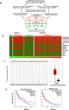
Fig. 1
Fig. 1. LINC00518 is overexpressed in CMM and indicates poor prognosis in patients with CMM.
A Schematic overview of the workflow used to identify and validate dysregulated lncRNAs in two CMM microarray data cohorts. B Heatmap of 12 upregulated probe sets representing 12 lncRNAs mined from the GSE4587 and GSE46517 data sets. C TCGA data showing that the Transcripts Per Million of LINC00518 in CMM was highest in all human cancers and that it is upregulated in CMM compared with normal tissues. D Kaplan–Meier curves of the OS and RFS of 436 patients with CMM with high or low LINC00518 expression. P value was computed by the log-rank test. SKCM stands for skin cutaneous melanoma.
Fig. 2
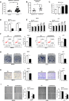
Fig. 2. LINC00518 knockdown suppressed cell proliferation, colony formation, migration and invasion, and reduced viability.
A The gene chip data related to LINC00518 were upregulated in T group (CMM tissues, n = 73) compared with N group in GSE46517 data set (normal tissues, n = 17). B qPCR assay showing that LINC00518 was overexpressed in 12 paired CMM tissues and their corresponding adjacent noncancerous skin tissues obtained from tissue biopsy samples in our study. C qPCR assay showing that LINC00518 expression was higher in WM451 and A375 cells, which have high invasive and metastatic tendency; however, it was lower in HM (normal melanocyte cell lines) and WM35 (low invasive and metastatic tendency) cells. The expression data in HM were considered as a control. D qPCR assay demonstrating shLINC00518 knockdown of LINC00518 in WM451 and A375 cell lines. E MTT assay indicating that LINC00518 knockdown in WM451/A375 cells decreased viability significantly compared with that in control cells. F Flow cytometry examination showing that LINC00518 knockdown could increase apoptosis of CMM cells. G Colony formation assay indicating that knockdown of LINC00518 could suppress cell proliferation in CMM cells. H Transwell assay showing the effect on CMM cell migration following LINC00518 knockdown. I Wound-healing assay showing that LINC00518 knockdown in WM451/A375 cells can significantly inhibit cell migration compared with the control. The histogram data for each group are an average of three independent replicates; bars indicate SD; *P < 0.05, **P < 0.01, ***P < 0.001.
Fig. 3
Fig. 3. LINC00518 directly targets miR-33a-3p and promotes HIF-1α expression.
A Kegg pathway analysis showed that LINC00518 may play a major role in glycolysis and TCA cycle. B Cytoscape was used to visualize LINC00518−miRNA−target gene interactions. The LINC00518/miRNAs interaction was predicted using RegRNA 2.0 and miRbase, the target genes of the miRNAs were predicted by combing GEO and TCGA databases with miRNA target prediction tools (microRNA.org and TargetScan). Red color demonstrates high expression level in CMM, and blue color demonstrates low expression level. C The expression levels of miR-33a-3p, miR-27a-3p, miR-18a-3p, miR216-5p, and miR-100-3p in melanoma cells following transfection with LINC00518 shRNA or NC. D The miR-33a-3p expression profile in human melanoma cell lines (WM35, WM451, A375) and human epidermal melanocytes (HM). E The HIF-1α expression profile in human melanoma cell lines (WM35, WM451, A375) and human epidermal melanocytes (HM). F Presumptive binding sites for miR-33a-3p were identified in the LINC00518 sequence and in the HIF-1α 3′ UTR by using sequence comparison and RegRNA data. G Negative correlation between LINC00518 and miR-33a-3p expressions in 12 cases of CMM tissues using Pearson correlation analysis. H RNA pull-down depicting that the bio-miR-33a-3p group was enriched for larger amounts of LINC00518 than the bio-miR-NC and bio-miR-33a-3p-MUT groups, thereby indicating that LINC00518 can target miR-33a-3p directly. I A dual-luciferase assay showing that miR-33a-3p mimics directly and significantly inhibited the relative dual-luciferase activity of the LINC00518-WT group compared with that of control group and LINC00518 targeted miR-33a-3p. Luciferase activity was detected 48 h after transfection. J qPCR assay showing a negative correlation between miR-33a-3p and HIF-1α expressions in 12 cases of CMM tissues. K Dual-luciferase assay of cells transfected with HIF-1α-3′UTR-WT or HIF-1α-3′UTR-MUT reporter together with miR-33a-3p mimic, LINC00518 shRNA, or LINC00518 shRNA plus miR-33a-3p inhibitor. L The changes of HIF-1α mRNA expression in WM451 and A375 transfected with miR-33a-3p mimic, LINC00518 shRNA, or LINC00518 shRNA plus miR-33a-3p inhibitor. M Western blotting analysis showing that HIF-1α and LDHA protein levels were regulated by LINC00518 and miR-33a-3p. The histogram data for each group are an average of three independent replicates; bars indicate SD; *P < 0.05, **P < 0.01, ***P < 0.001.
Fig. 4
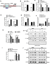
Fig. 4. HIF-1α negatively regulates miR-33a-3p expression in CMM cells.
A JASPAR predicted the binding sites of the HIF-1α and LINC00518 promoter regions. B qRT-PCR showing that the expression of miR-33a-3p was significantly increased after HIF-1α knockout. C, D The expression changes of miR-33a-3p and LINC00518 after transfected with shHIF-1α, shLINC00518 or shLINC00518 plus HIF-1α overexpression. E Dual-luciferase assay showing the luciferase activity in HEK293T cells cotransfected with wild-type (Wt) or mutant (Mut) HIF-1α in the miR-33a-3p promoter region. F ChIP PCR indicated that the miR-33a-3p promoter region bound to HIF-1α. G qRT-PCR showing miR-33a-3p expression levels after HDAC1, HDAC2, HDAC3 knockout in CMM cells. H CoIP assay showed that the HIF-1α-HDAC immunoprecipitate was decreased after HIF-1α knockdown and increased after HIF-1α overexpression. I ChIP PCR showing that Santacruzamate A (inhibiting histone deacetylation) could recover the relative expression levels of miR-33a-3p in WM451 and A375 cells; immunoglobulin G (IgG) acted as the NC. J Western blotting analysis, indicating that LINC00518 knockdown decreased HIF-1α and LDHA protein levels, while overexpression of HIF-1α in LINC00518 knockdown cells enhanced HIF-1α and LDHA expression. The histogram data for each group are an average of three independent replicates; bars indicate SD; *P < 0.05, **P < 0.01, ***P < 0.001.
Fig. 5
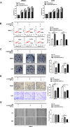
Fig. 5. Overexpression of HIF-1α reversed LINC00518 knockdown-induced suppressing of proliferation, migration, invasion, and colony formation.
A WM451 and A375 cells were transfected with NC, shLINC00518, shLINC00518 plus HIF-1α overexpression plasmid; the MTT method was used to detect cell proliferation. B The effects of LINC00518 knockdown or LINC00518 knockdown plus HIF-1α overexpression on apoptosis of WM451 or A375 cells were assessed using annexin V staining and flow cytometry analysis. C Colony formation assay showed that LINC00518 silenced is involved in significantly decreasing cell proliferation, HIF-1α overexpression could reverse the regulatory effects of LINC00518. D Invasion of melanoma cells in different transfection groups was assessed by the transwell assay. E Wound-healing assay was used to assess the migration of melanoma cells in different transfection groups. The histogram data for each group are an average of three independent replicates; bars indicate SD; *P < 0.05, **P < 0.01, ***P < 0.001.
Fig. 6
Fig. 6. Involvement of the LINC00518–miR-33a-3pHIF-1α negative feedback loop in glycolysis-mediated radiotherapy resistance of CMM cells.
AC The levels of glucose consumption and lactate production were estimated in CMM cells. These results indicated that knockdown of LINC00518 expression in WM451 and A375 cells decreased glucose consumption, ATP levels, and lactic acid production in the cells; by contrast, overexpression of HIF-1α significantly reversed the effects of LINC00518 silencing on glycolytic tumor metabolism in WM451 and A375 cells. D Detection of CMM cell sensitivity to radiotherapy after LINC00518 knockdown by using an MTT assay. LINC00518 knockdown in CMM cells could inhibit their proliferation after the use of radiotherapy and HIF-1α could reverse this effect, while 2DG and Santacruzamate A offset the influence of HIF-1α. E Flow cytometry examination of the effect of radiotherapy on CMM cell apoptosis following LINC00518 knockdown. LINC00518 knockdown in CMM cells could increase apoptosis after using radiotherapy and HIF-1α could reverse this effect, while 2DG and Santacruzamate A offset the influence of HIF-1α. The histogram data for each group are an average of three independent replicates; bars indicate SD; *P < 0.05, **P < 0.01, ***P < 0.001.
Fig. 7
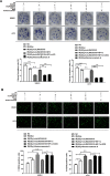
Fig. 7. Involvement of the LINC00518/miR-33a-3p/HIF-1α negative feedback loop in glycolysis-mediated radiotherapy resistance of CMM cells.
A Colony formation showing that knockdown of LINC00518 expression in WM451 and A375 cells inhibited the invasion and proliferation of CMM cells, increased the radiosensitivity of cells, while overexpression of HIF-1α reversed this effect. Both 2DG and Santacruzamate A increased radiosensitivity, which was inhibited by HIF-1α overexpression or by upregulation of LINC00518 and HIF-1α in the CMM cells. B Immunofluorescence assays indicating that knockdown of LINC00518 expression in WM451 and A375 cells could induce structural damage of their nuclei under radiotherapy; however, HIF-1α could reverse this effect, while 2DG and Santacruzamate A offset the influence of HIF-1α. The histogram data for each group are an average of three independent replicates; bars indicate SD; *P < 0.05, **P < 0.01, ***P < 0.001.
Fig. 8
Fig. 8. Effect of the LINC00518/miR-33a-3p/HIF-1α negative feedback loop on radiosensitivity in CMM cells in vivo.
A Transplantation tumor experiments showing that repression of LINC00518 expression using the shLINC00518 plasmid or treating the cells with Santacruzamate A reduced the tumorigenic ability and tumor volume of WM451 and A375 cells, thereby increasing their radiosensitivity as observed by reduced colony formation in the subcutaneous sarcoma model. B Immunohistochemical assays showing that knockdown of LINC00518 expression or treating the cells with Santacruzamate A decreased HIF-1α, Ki67, and LDHA protein levels in tumor tissues of mice. C Western blotting showing that LINC00518 expression or treatment of the cells with Santacruzamate A decreased HIF-1α and LDHA protein expression in tumor tissues of mice. D Schematic of this search. The histogram data for each group are an average of three independent replicates; bars indicate SD; *P < 0.05, **P < 0.01, ***P < 0.001.

Similar articles

Cited by

References

    1. Schadendorf D, et al. Melanoma. Nat. Rev. Dis. 2015;1:1–20. - PubMed
    1. Rastrelli M, Tropea S, Rossi C, Alaibac M. Melanoma: epidemiology, risk factors, pathogenesis, diagnosis and classification. In Vivo. 2014;28:1005–1011. - PubMed
    1. Lee C, Thoma C, Ng K. An overview of the changing landscape of treatment for advanced melanoma. Pharmacotherapy. 2017;37:319–333. doi: 10.1002/phar.1895. - DOI - PubMed
    1. Eggermont A, Spatz A, Robert C. Cutaneous melanoma. Lancet. 2014;383:816–827. doi: 10.1016/S0140-6736(13)60802-8. - DOI - PubMed
    1. Balch C, Gershenwald J, Soong S, Thompson J, Atkins M. Final version of 2009 AJCC melanoma staging and classification. J. Clin. Oncol. 2009;27:199–6206. doi: 10.1200/JCO.2009.23.4799. - DOI - PMC - PubMed

Publication types

MeSH terms

Substances