. 2021 Mar 4;12(1):1443.
doi: 10.1038/s41467-021-21663-w.

The methyltransferase SETD2 couples transcription and splicing by engaging mRNA processing factors through its SHI domain

Affiliations

The methyltransferase SETD2 couples transcription and splicing by engaging mRNA processing factors through its SHI domain

Saikat Bhattacharya et al. Nat Commun. .

Abstract

Heterogeneous ribonucleoproteins (hnRNPs) are RNA binding molecules that are involved in key processes such as RNA splicing and transcription. One such hnRNP protein, hnRNP L, regulates alternative splicing (AS) by binding to pre-mRNA transcripts. However, it is unclear what factors contribute to hnRNP L-regulated AS events. Using proteomic approaches, we identified several key factors that co-purify with hnRNP L. We demonstrate that one such factor, the histone methyltransferase SETD2, specifically interacts with hnRNP L in vitro and in vivo. This interaction occurs through a previously uncharacterized domain in SETD2, the SETD2-hnRNP Interaction (SHI) domain, the deletion of which, leads to a reduced H3K36me3 deposition. Functionally, SETD2 regulates a subset of hnRNP L-targeted AS events. Our findings demonstrate that SETD2, by interacting with Pol II as well as hnRNP L, can mediate the crosstalk between the transcription and the splicing machinery.


Conflict of interest statement

The authors declare no competing interests.

Figures

Fig. 1
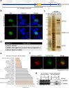
Fig. 1. Purification of hnRNP L RRM2 reveals SETD2 as an interactor.
a Cartoon illustrating the truncation of hnRNP L along with the known domains. RRM RNA-recognition motif, NLS nuclear localization signal. b Microscopy images showing localization of mCherry-hnRNP L 162–321. The scale bar is 10 µm. The experiment was repeated at least four times all yielding similar results. c, d Halo purification was performed from extracts of 293T cells expressing Halo-hnRNP L 162–321. Input and eluted samples were resolved on gel followed by silver staining or western blotting. The expected band for the target proteins are depicted by arrows. HE high exposure, LE low exposure. The experiment was repeated at least two times all yielding similar results. e IPA (Ingenuity Pathway Analysis) of proteins enriched in Halo-hnRNP L 162–321 purification. f, g Table showing the dNSAFs ×100 of the listed protein. dNSAF distributed normalized spectral abundance factor.
Fig. 2
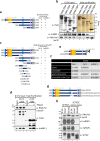
Fig. 2. SETD2 reciprocally co-purifies hnRNP L.
a Cartoon illustrating the overlapping segments of SETD2 used for affinity purification along with the known domains. AWS associated with SET, SET Su(var)3–9, Enhancer-of-zeste and Trithorax, SRI Set2-Rpb1 interaction. b Microscopy images showing localization of GFP-SETD2 fragments. The scale bar is 10 µm. The experiment was repeated at least eight times all yielding similar results. c Halo purification was performed from extracts of 293T cells expressing Halo-SETD2C. Input and eluted samples were resolved on a gel followed by silver staining. M—protein marker. The experiment was repeated at least 10 times all yielding similar results. d Table showing the dNSAFs (distributed normalized spectral abundance factor) of the listed proteins. e IPA (Ingenuity Pathway Analysis) of proteins enriched in Halo-SETD2C purification. AP-MS affinity purification-mass spectrometry. f Microscopy images showing localization of mCherry-hnRNP L and GFP-SETD2C fragment. The scale bar is 10 µm. GFP green fluorescent protein. The experiment was repeated at least four times all yielding similar results. g Halo purification was performed from extracts of 293T cells expressing Halo-SETD2C. Input and eluted samples were resolved on a gel and probed with an anti-hnRNP L antibody. The experiment was repeated at least two times all yielding similar results.
Fig. 3
Fig. 3. Domain mapping experiment reveals a novel SETD2-hnRNP interaction (SHI) domain in SETD2.
RNase treatment was not performed for these experiments. a, c, e, g Cartoon illustrating the SETD2 and ySet2 constructs along with the known domains that were used in affinity-purifications. b, d, h Halo purification was performed from extracts of 293T cells expressing Halo- or Halo-FLAG-tagged proteins. Input and eluted samples were resolved on gel followed by silver staining or western blotting. The expected band for the target proteins are depicted by arrows. HE high exposure, LE low exposure, * non-specific. The experiments were repeated at least two times all yielding similar results. f Table showing the dNSAFs of the listed proteins post mass spectrometry analysis of purified complexes obtained by affinity purification of Halo-Set2 from 239T extracts. AWS associated with SET, SET Su(var)3–9, Enhancer-of-zeste and Trithorax, SRI Set2-Rpb1 Interaction, dNSAF distributed normalized spectral abundance factor, NLS nuclear localization signal.
Fig. 4
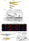
Fig. 4. SETD2 and hnRNP L interact in vitro.
a, d Cartoon illustrating the hnRNP L and SETD2 constructs along with the known domains that were used in affinity-purifications and in vitro binding. b Halo purification was performed from extracts of 293T cells co-expressing Halo-tagged SETD2C and mCherry-HA-hnRNP L. Input and eluted samples were resolved on gel followed by western blotting. The expected band for the target proteins are depicted by arrows. RNase treatment was not performed for these experiments. The experiment was repeated at least two times all yielding similar results. c Microscopy images showing the localization of mCherry-hnRNP L constructs. The scale bar is 10 µm. The experiment was repeated at least four times all yielding similar results. e GST pull-down was performed using recombinant proteins purified from bacteria. RNase was included in the binding assay. The input and eluted samples were resolved on gel followed by western blotting with the depicted antibodies. The experiment was repeated at least two times all yielding similar results. AWS associated with SET, SET Su(var)3–9, Enhancer-of-zeste and Trithorax, SRI Set2-Rpb1 interaction, SHI SETD2-hnRNP interaction, RRM RNA-recognition motif, NLS nuclear localization signal, GST glutathione-S-transferase.
Fig. 5
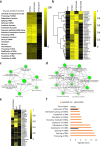
Fig. 5. SETD2 associates with splicing-related proteins through its SHI domain.
a, b, e Heat maps showing the enrichment of pathways in the IPA (Ingenuity Pathway Analysis) and proteins in MudPIT analysis. c, d GO-term analysis of proteins using ShinyGO (http://bioinformatics.sdstate.edu/go/) identified by MudPIT in the affinity purification of SETD2 SHI and 1964–2113 fragments. f Chart showing the enriched pathways in IPA of ySet2 proteins. SRI Set2-Rpb1 interaction, SHI SETD2-hnRNP interaction.
Fig. 6
Fig. 6. SETD2 and hnRNP L depletion affect transcription and splicing of a common subset of genes.
a RNA was isolated from 293T cells transfected with siRNA and RTPCR was performed to check transcript levels. gapdh was used as a normalization control. Western blot of whole-cell lysates was performed with the depicted antibodies. The experiment was repeated at least seven times all yielding similar results. b Chart showing the decrease in expression of the genes depicted based on RNA-seq analysis post siRNA treatment. c, e, f Pie charts showing the fractions of differentially expressed genes and AS events that occur in both setd2 and hnrnpl depletion. d Heat map showing the genes that show differential expression in both setd2 and hnrnpl depleted 293T cells. AS alternative splicing, A3SS alternate 3′ splice site, A5SS alternate 5′ splice site, RI retained intron, MXE mutually exclusive exon, SE skipped exon.
Fig. 7
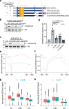
Fig. 7. The SHI domain regulates SETD2 activity.
a Cartoon illustrating the SETD2 constructs along with their known domains that were used to compare the ability to deposit H3K36me3 in KO cells. b, c Western blot with the depicted antibodies of whole-cell lysates of KO cells expressing SETD2 mutants. The experiment was repeated at least five times all yielding similar results. d Bar graph showing H3 normalized H3K36me3 signal intensity of data depicted in (c). n = 4 independent biological samples examined in four independent experiments. Unpaired t test (two-tailed) was performed. p-value <0.05 (FLΔSRI vs FL = 0.0071; FLΔSHI vs FL = 0.0344; FLΔSRIΔSHI vs FL = 0.0013) was considered significant. Data are presented as mean values with standard error of mean. e, f, g, h Metagene plot and boxplot depicting the distribution of H3K36me3 upon expression of SETD2 FL, FLΔSRI, and FLΔSHI in setd2Δ 293T cells. For each sample n = 2 independent biological samples examined in the same sequencing run. In the boxplots, the black line inside the box shows the median. The box bottom and top border correspond to 25th and 75th percentiles (Q1 and Q3, respectively). The whiskers represent ranges from Q1 − 1.5 * IQR to Q3 + 1.5 * IQR where IQR stands for interquartile range (Q3–Q1). Data points outside the whiskers could be outliers and are marked as black dots. AWS associated with SET, SET Su(var)3–9, Enhancer-of-zeste and Trithorax, SRI Set2-Rpb1 interaction, SHI SETD2-hnRNP interaction, NLS nuclear localization signal, KO knock out (setd2Δ 293T cells), TSS transcription start site, TES transcription end site.
Fig. 8
Fig. 8. Pre-mRNA processing and transcription are coupled.
A cartoon speculating a possible mechanism of cross talk between hnRNP L and the transcription machinery. See text for details.

References

    1. Kelemen O, et al. Function of alternative splicing. Gene. 2013;514:1–30. doi: 10.1016/j.gene.2012.07.083. - DOI - PMC - PubMed
    1. Pan Q, Shai O, Lee LJ, Frey BJ, Blencowe BJ. Deep surveying of alternative splicing complexity in the human transcriptome by high-throughput sequencing. Nat. Genet. 2008;40:1413–1415. doi: 10.1038/ng.259. - DOI - PubMed
    1. Wang ET, et al. Alternative isoform regulation in human tissue transcriptomes. Nature. 2008;456:470–476. doi: 10.1038/nature07509. - DOI - PMC - PubMed
    1. Barbosa-Morais NL, et al. The evolutionary landscape of alternative splicing in vertebrate species. Science. 2012;338:1587. doi: 10.1126/science.1230612. - DOI - PubMed
    1. Kalsotra A, Cooper TA. Functional consequences of developmentally regulated alternative splicing. Nat. Rev. Genet. 2011;12:715–729. doi: 10.1038/nrg3052. - DOI - PMC - PubMed

Publication types

MeSH terms