

Specific microstructural changes of the cervical spinal cord in syringomyelia estimated by diffusion tensor imaging
- PMID: 33664296
- PMCID: PMC7933234
- DOI: 10.1038/s41598-021-84164-2
Specific microstructural changes of the cervical spinal cord in syringomyelia estimated by diffusion tensor imaging
Abstract
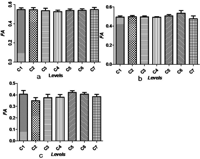
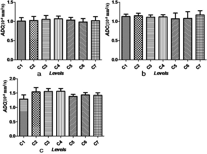
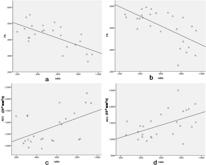
The microstructure of the spinal cord in syringomyelia has not been well studied. The aim of this study was to evaluate the microstructure of the cervical cord in patients with syringomyelia using diffusion tensor imaging (DTI) and to investigate the association between DTI parameters and the size of the syrinx cavity. Thirty patients with syringomyelia and 11 age-matched controls were included in this study. DTI and T1/T2-weighted MRI were used to estimate spinal microstructure. The patients were divided into a clinical symptom group (group A) and a non-clinical symptom group (group B) according to ASIA assessments. The fractional anisotropy (FA) and apparent diffusion coefficient (ADC) values (mm2/s) were measured and compared between patients and controls. Correlation between FA/ADC and the size of the syrinx cavity was examined with a bivariate analysis. FA values were lower (P < 0.000) and ADC values were higher (P < 0.000) compared to the controls at the level of all syrinxes examined in patients with syringomyelia; both FA values and ADC values reached normal values either above or below the syrinx levels (all P > 0.05). FA values and ADC values at all cervical levels were not significantly different either in controls or outside of the syrinx (all P > 0.05). FA values of group A was significantly lower than those of group B (P < 0.000). There was a negative association between FA values and the size of syrinx cavity, and a positive association between ADC values and the size of syrinx cavity (FA: P < 0.05, ADC: P < 0.05). The microstructure of the cervical spinal cord is different across all patients with syringomyelia. DTI is a promising tool for estimating quantitative pathological characteristics that are not visible with general MRI.
Conflict of interest statement
The authors declare no competing interests.
Figures


Similar articles
-
Changes and clinical correlation of diffusion tensor imaging parameters of compressed spinal cord and nerve root in patients with cervical spondylosis.BMC Med Imaging. 2022 Jun 3;22(1):107. doi: 10.1186/s12880-022-00835-0. BMC Med Imaging. 2022. PMID: 35659198 Free PMC article.
-
Diffusion tensor imaging in cervical syringomyelia secondary to Chiari I malformation: preliminary results.Spine (Phila Pa 1976). 2015 Apr 1;40(7):E381-7. doi: 10.1097/BRS.0000000000000781. Spine (Phila Pa 1976). 2015. PMID: 25584946
-
Correlation of magnetic resonance diffusion tensor imaging and clinical findings of cervical myelopathy.Spine J. 2013 Aug;13(8):867-76. doi: 10.1016/j.spinee.2013.02.005. Epub 2013 Mar 21. Spine J. 2013. PMID: 23523441
-
A meta-analysis of the role of diffusion tensor imaging in cervical spinal cord compression.J Neuroimaging. 2023 Jul-Aug;33(4):493-500. doi: 10.1111/jon.13093. Epub 2023 Mar 13. J Neuroimaging. 2023. PMID: 36914383 Review.
-
The role of diffusion tensor imaging in the diagnosis, prognosis, and assessment of recovery and treatment of spinal cord injury: a systematic review.Neurosurg Focus. 2019 Mar 1;46(3):E7. doi: 10.3171/2019.1.FOCUS18591. Neurosurg Focus. 2019. PMID: 30835681
Cited by
-
Diffusion Tensor Imaging in Syringomyelia Secondary to Chiari Malformation in Cavalier King Charles Spaniel-A Preliminary Study.Animals (Basel). 2022 Dec 2;12(23):3405. doi: 10.3390/ani12233405. Animals (Basel). 2022. PMID: 36496926 Free PMC article.
-
Identification of Atypical Scoliosis Patterns Using X-ray Images Based on Fine-Grained Techniques in Deep Learning.Global Spine J. 2025 Jun 11:21925682251349999. doi: 10.1177/21925682251349999. Online ahead of print. Global Spine J. 2025. PMID: 40500925 Free PMC article.
-
Changes and clinical correlation of diffusion tensor imaging parameters of compressed spinal cord and nerve root in patients with cervical spondylosis.BMC Med Imaging. 2022 Jun 3;22(1):107. doi: 10.1186/s12880-022-00835-0. BMC Med Imaging. 2022. PMID: 35659198 Free PMC article.
-
Predictive value of dynamic diffusion tensor imaging for surgical outcomes in patients with cervical spondylotic myelopathy.BMC Med Imaging. 2024 Oct 1;24(1):260. doi: 10.1186/s12880-024-01428-9. BMC Med Imaging. 2024. PMID: 39354411 Free PMC article.
References
-
- Vandertop WP. Syringomyelia. Neuropediatrics. 2014;45:3–9. - PubMed
-
- Hale, A. T. et al. Factors associated with syrinx size in pediatric patients treated for Chiari malformation type I and syringomyelia: a study from the Park-Reeves syringomyelia research consortium. J. Neurosurg. Pediatr. 10.3171/2020.1.PEDS19493. 2020. - PubMed
-
- Tohyama, S., Walker, M. R., Sammartino, F., Krishna, V. & Hodaie, M. The utility of diffusion tensor imaging in neuromodulation: moving beyond conventional magnetic resonance imaging. Neuromodulation. 23, 427–435 (2020). - PubMed
-
- Derek KJ, Thomas RK, Robert T. White matter integrity, fiber count, and other fallacies: the do's and don'ts of diffusion MRI. Neuroimage. 2019;73:239–254. - PubMed
Publication types
MeSH terms
LinkOut - more resources
Full Text Sources
Other Literature Sources
Medical
