. 2021 Mar 4;11(1):5139.
doi: 10.1038/s41598-021-84230-9.

Ambient noise exposure induces long-term adaptations in adult brainstem neurons

Affiliations

Ambient noise exposure induces long-term adaptations in adult brainstem neurons

Ida Siveke et al. Sci Rep. .

Abstract

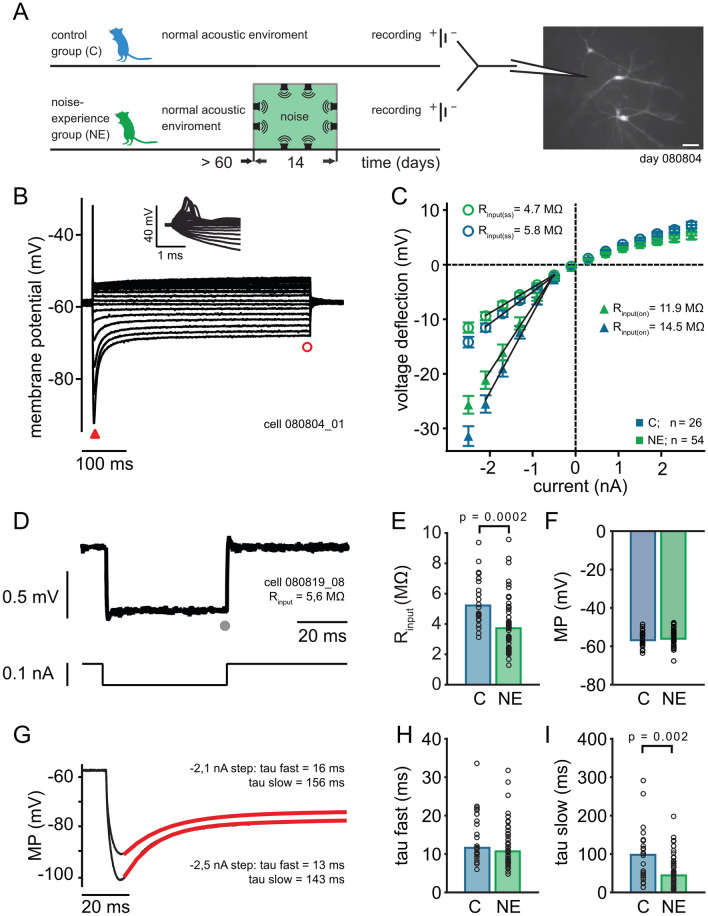
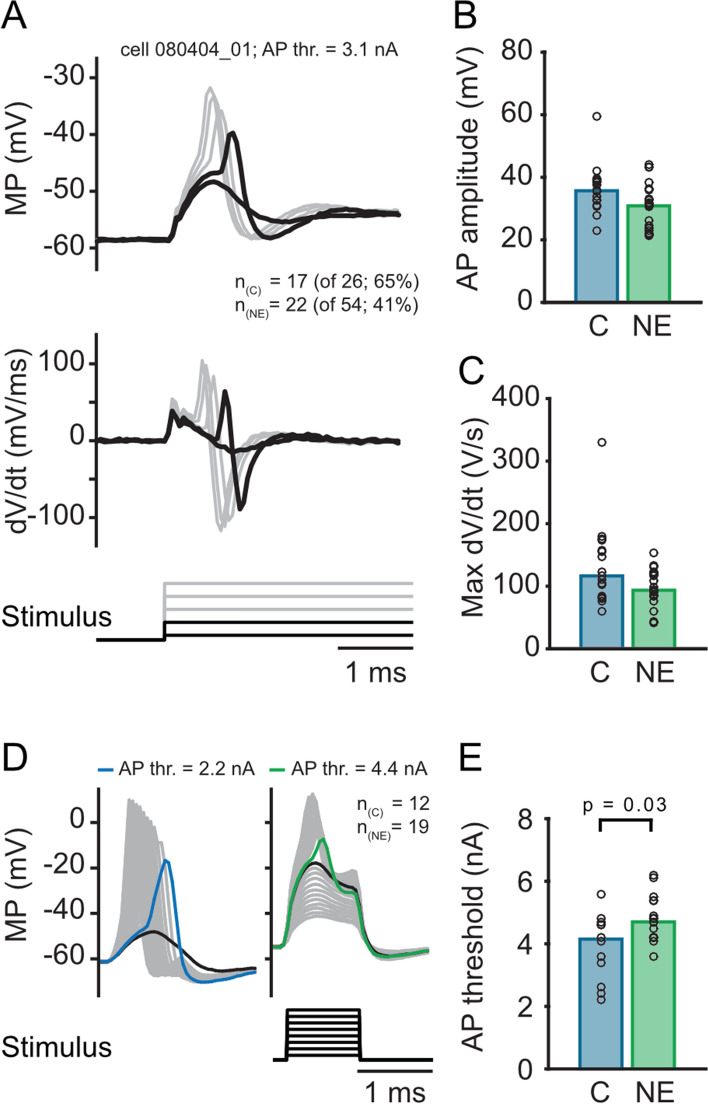
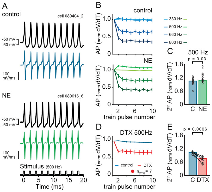
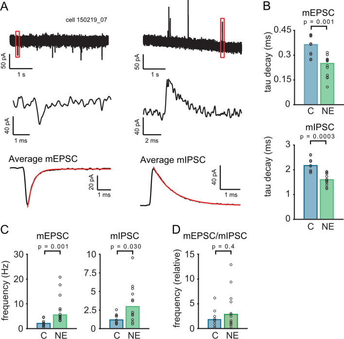
To counterbalance long-term environmental changes, neuronal circuits adapt the processing of sensory information. In the auditory system, ongoing background noise drives long-lasting adaptive mechanism in binaural coincidence detector neurons in the superior olive. However, the compensatory cellular mechanisms of the binaural neurons in the medial superior olive (MSO) to long-term background changes are unexplored. Here we investigated the cellular properties of MSO neurons during long-lasting adaptations induced by moderate omnidirectional noise exposure. After noise exposure, the input resistance of MSO neurons of mature Mongolian gerbils was reduced, likely due to an upregulation of hyperpolarisation-activated cation and low voltage-activated potassium currents. Functionally, the long-lasting adaptations increased the action potential current threshold and facilitated high frequency output generation. Noise exposure accelerated the occurrence of spontaneous postsynaptic currents. Together, our data suggest that cellular adaptations in coincidence detector neurons of the MSO to continuous noise exposure likely increase the sensitivity to differences in sound pressure levels.


Conflict of interest statement

The authors declare no competing interests.

Figures

Figure 1
Figure 1
Intrinsic properties of MSO principal neurons from adult animals with and without noise exposure: neurons are getting leaky and faster after noise exposure. (A) Schematic drawing of the experimental design and three typical principal MSO neurons filled with Alexa Fluor 568 during recording (right picture). (B) Response to different 500 ms current pulses (− 2.5 to 4.3 nA; 0.4 nA steps) using current clamp recordings while blocking synaptic transmission (SR—GABA antagonist, D-AP5-NMDA antagonist, TTX – Na1). (C), On-current (on; triangle, see (B) for example) and the steady-state-current (ss; circle, see (B)) V–I plots generated from the average voltage responses of ≥ 3 repetitions. The average Rinput was estimated from the slope of the V–I plots between − 2.1 and − 0.5 nA current application. (D) Response to a small current injection (− 0.1 nA, grey lines) were used to estimate the steady state (grey dot) Rinput of each cell to the average response (black line; ≥ 20 repetitions). (E) The Rinput of the control neurons (median = 5.2 MΩ) is significant higher (p = 0.0002) as the resistance of neurons of animals exposed to noise (median = 3.7 MΩ). (F) The membrane potentials (MP) do not differ between the two groups. (G) Example average response (≥ 3 repetitions) to high negative currents (− 2.5 and − 2.1 nA) were used to estimate the membrane potential and the time constants of the depolarizing sag (defined by a double exponential fit, red lines) during negative current injections. (H) The fast time constants of the depolarizing sag potential were not changed during noise exposure (c = 11.6 ms; NE = 10.7 ms). (I) The slow time constants were significant smaller (p = 0.002) in neurons of noise exposed animals (c = 0.098 s; NE = 0.044 s). Bars represent the median values, significance was assessed using two-sided Wilcoxon rank sum test.
Figure 2
Figure 2
Action potential characteristics and generation in MSO neurons from adult animals with and without noise exposure: Action potentials are getting smaller and faster, and the threshold to generate action potential is higher after noise exposure. (A) Example action potentials of a principal MSO neuron induced by high currents (see Fig. 1B). The maximal current of 2.4 nA induced action potential in 65% (nc = 17 of 26 tested) of the neurons of the control group but only in 41% (nNE 22 of 54 tested) neurons of the NE group. The upper graph shows an exemplary recording in mV, the lower graph the first derivative (dV/dt). (B) Amplitudes of the action potentials (AP) (c = 35.7 mV; NE = 30.9 mV, p = 0.040, without outlier/grey dot p = 0.069) and slope of the action potentials (C) (expressed in dV/dt) (c = 116.5 V/s; NE = 93.6 V/s, p = 0.087, without outlier grey dot p = 0.14) of the two groups. (D) Example action potentials evoked by a family of short increasing 1 ms currents (0.1 to 7 nA, 0.1nA steps). This stimulus was used in a subset of MSO neurons to precisely investigate the action potential threshold. (E) Neurons that elicited an action potential by a maximal current of 7 nA, showed a significantly higher action potential threshold (c = 4.2 nA; NE = 4.7 nA, p = 0.03) in NE neurons. Bars represent the median values, significance was assessed using two-sided Wilcoxon rank sum test.
Figure 3
Figure 3
Action potential generation is facilitated at higher frequencies after noise exposure. (A) Exemplary raw traces (three repetition) and the first derivative (dV/dt) of the response to a train of 10 stimuli (indicated below). The upper panel shows the data of the control group the lower panel the data of the NE group. (B) Mean response of action potential (AP) depolarisation speed (dV/dt) (± SEM normalized to the first action potential of the 10-stimuli train shown for four different frequencies (the control group: upper panel, NE group: bottom panel). (C) The rise of the second amplitude is significantly increased at the 500 Hz-train after noise exposure (p = 0.0348). (D) Mean action potential depolarisation speed (dV/dt) (± SEM) after blocking the Kv1 channel with DTX (Dendrotoxin 100 nM) for the 10 pulse 500 Hz-stimulus train. (E) Action potential depolarisation speed decreases significantly (p = 0.0006) in neurons after DTX application. Bars represent the median values, significance was assessed using two-sided Wilcoxon’s signed rank (E) and two-sided Wilcoxon rank sum test (C).
Figure 4
Figure 4
Characterization of miniature EPSCs and IPSCs in MSO neurons from adult animals with and without noise exposure: While the amplitude of the miniature events stays constant, the events are faster and their frequency is increased. (A) Recording of mEPSCs (left) and mIPSCs (right) in the same neuron using different holding potentials (− 60 mV for mEPCSs and + 10 mV for mIPSCs). The middle graph shows a single enlarged mEPSC or mIPSC from the recording (upper graph, red box) and the bottom graph the average mEPCS or mIPSC. (B) Decay time constants (tau decay) of the average mEPSCs (p = 0.0014) and mIPSCs (p = 0.0003). (C) Frequency of mEPSCs (left; p = 0.00095) and mIPSCs (right; p = 0.0302) significantly increases in neurons after noise exposure. (D) The relative frequency (left; p = 0.414) of mEPSCs/mIPSCs does not change after noise exposure.

Similar articles

Cited by

References

    1. Keating P, King AJ. Sound localization in a changing world. Curr. Opin. Neurobiol. 2015;35:35–43. doi: 10.1016/j.conb.2015.06.005. - DOI - PubMed
    1. Keuroghlian AS, Knudsen EI. Adaptive auditory plasticity in developing and adult animals. Prog. Neurobiol. 2007;82:109–121. doi: 10.1016/j.pneurobio.2007.03.005. - DOI - PubMed
    1. Rabinowitz NC, Willmore BD, King AJ, Schnupp JW. Constructing noise-invariant representations of sound in the auditory pathway. PLoS Biol. 2013;11:e1001710. doi: 10.1371/journal.pbio.1001710. - DOI - PMC - PubMed
    1. Wark B, Lundstrom BN, Fairhall A. Sensory adaptation. Curr. Opin. Neurobiol. 2007;17:423–429. doi: 10.1016/j.conb.2007.07.001. - DOI - PMC - PubMed
    1. Illing RB. Activity-dependent plasticity in the adult auditory brainstem. Audiol. Neurootol. 2001;6:319–345. doi: 10.1159/000046844. - DOI - PubMed

Publication types

MeSH terms

LinkOut - more resources