

Peripheral ERK modulates acupuncture-induced brain neural activity and its functional connectivity
- PMID: 33664320
- PMCID: PMC7933175
- DOI: 10.1038/s41598-021-84273-y
Peripheral ERK modulates acupuncture-induced brain neural activity and its functional connectivity
Abstract
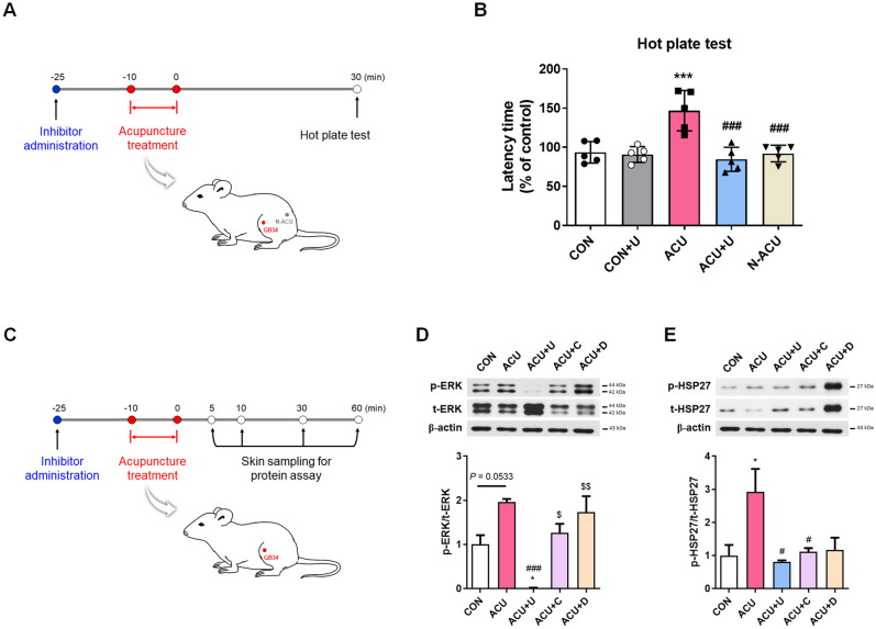
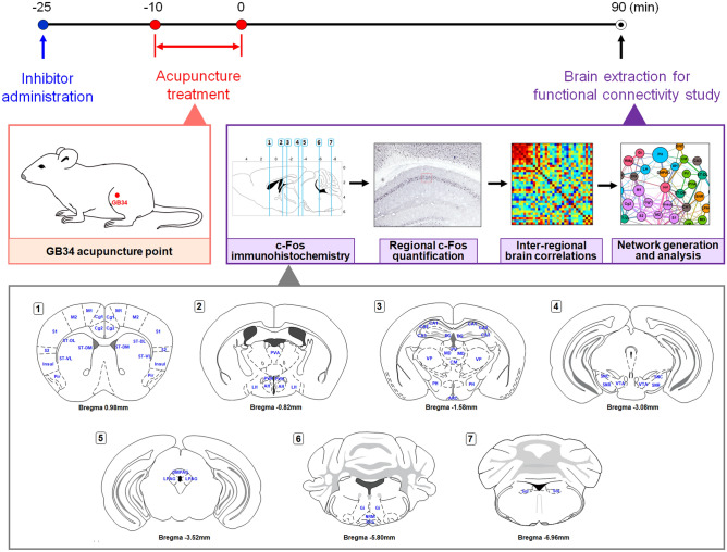
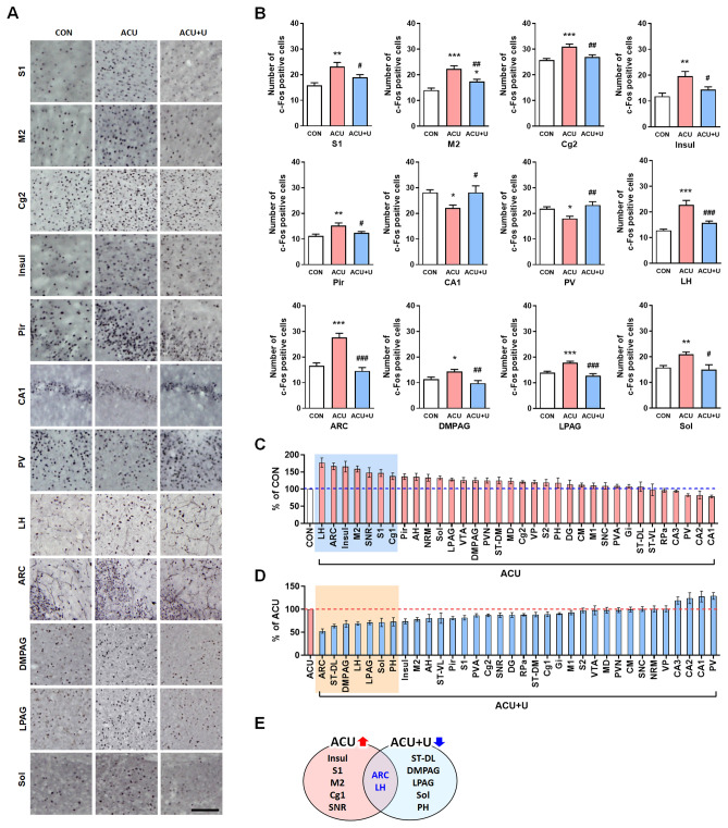
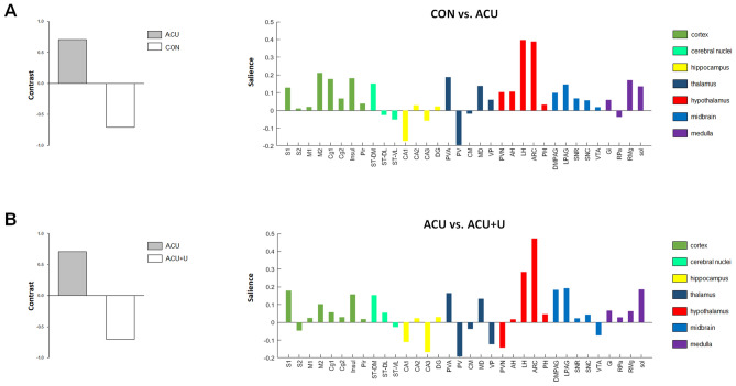
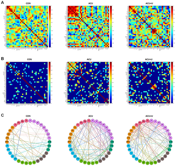
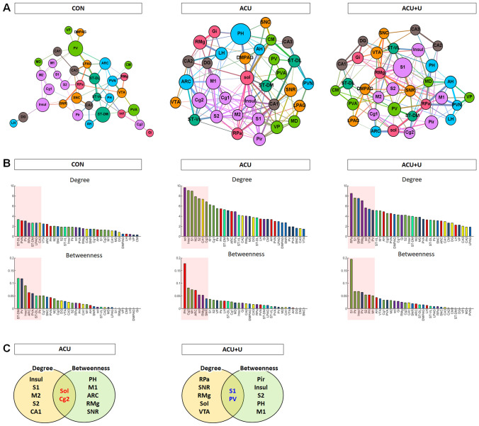
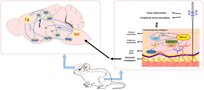
Acupuncture has been widely used as a therapeutic intervention, and the brain network plays a crucial role in its neural mechanism. This study aimed to investigate the acupuncture mechanism from peripheral to central by identifying how the peripheral molecular signals induced by acupuncture affect the brain neural responses and its functional connectivity. We confirmed that peripheral ERK activation by acupuncture plays a role in initiating acupuncture-induced peripheral proteomic changes in mice. The brain neural activities in the neocortex, hippocampus, thalamus, hypothalamus, periaqueductal grey, and nucleus of the solitary tract (Sol) were significantly changed after acupuncture, and these were altered by peripheral MEK/MAPK inhibition. The arcuate nucleus and lateral hypothalamus were the most affected by acupuncture and peripheral MEK/MAPK inhibition. The hypothalamic area was the most contributing brain region in contrast task PLS analysis. Acupuncture provoked extensive changes in brain functional connectivity, and the posterior hypothalamus showed the highest betweenness centrality after acupuncture. After brain hub identification, the Sol and cingulate cortex were selected as hub regions that reflect both degree and betweenness centrality after acupuncture. These results suggest that acupuncture activates brain functional connectivity and that peripheral ERK induced by acupuncture plays a role in initiating brain neural activation and its functional connectivity.
Conflict of interest statement
The authors declare no competing interests.
Figures
Similar articles
-
[Effect of acupuncture on hypothalamic functional connectivity in patients with premature ova-rian insufficiency based on resting-state functional magnetic resonance imaging].Zhen Ci Yan Jiu. 2022 Jul 25;47(7):617-24. doi: 10.13702/j.1000-0607.20210399. Zhen Ci Yan Jiu. 2022. PMID: 35880279 Chinese.
-
Modulatory Effects of Actual and Imagined Acupuncture on the Functional Connectivity of the Periaqueductal Gray and Ventral Tegmental Area.Psychosom Med. 2021 Oct 1;83(8):870-879. doi: 10.1097/PSY.0000000000000984. Psychosom Med. 2021. PMID: 34292206 Free PMC article. Clinical Trial.
-
[Effect of acupuncture on pain-emotion related brain regions in patients with cervical spondylosis of cervical type: a fMRI study].Zhongguo Zhen Jiu. 2021 Aug 12;41(8):906-12. doi: 10.13703/j.0255-2930.20201022-k0007. Zhongguo Zhen Jiu. 2021. PMID: 34369703 Chinese.
-
Research progress of acupuncture regulating ERK signaling pathway in the treatment of depression.Zhen Ci Yan Jiu. 2024 May 25;49(5):519-525. doi: 10.13702/j.1000-0607.20230278. Zhen Ci Yan Jiu. 2024. PMID: 38764124 Review. Chinese, English.
-
Acupuncture-induced pain relief and the human brain's default mode network - an extended view of central effects of acupuncture analgesia.Forsch Komplementmed. 2012;19(4):197-201. doi: 10.1159/000341928. Epub 2012 Aug 3. Forsch Komplementmed. 2012. PMID: 22964986 Review.
Cited by
-
Peripheral Rho-associated protein kinase activation mediates acupuncture analgesia.Integr Med Res. 2024 Sep;13(3):101051. doi: 10.1016/j.imr.2024.101051. Epub 2024 May 31. Integr Med Res. 2024. PMID: 39219984 Free PMC article.
-
Network Neuroscience Untethered: Brain-Wide Immediate Early Gene Expression for the Analysis of Functional Connectivity in Freely Behaving Animals.Biology (Basel). 2022 Dec 24;12(1):34. doi: 10.3390/biology12010034. Biology (Basel). 2022. PMID: 36671727 Free PMC article. Review.
-
Abnormal thalamocortical network dynamics in patients with migraine and its relationship with electroacupuncture treatment response.Brain Imaging Behav. 2024 Dec;18(6):1467-1479. doi: 10.1007/s11682-024-00938-y. Epub 2024 Sep 28. Brain Imaging Behav. 2024. PMID: 39340626
-
Acupuncture for treating attention deficit hyperactivity disorder in children: A protocol for systematic review and meta-analysis.PLoS One. 2022 Oct 10;17(10):e0275504. doi: 10.1371/journal.pone.0275504. eCollection 2022. PLoS One. 2022. PMID: 36215241 Free PMC article.
-
The Involvement of the Ventral Tegmental Area in the Electroacupuncture Alleviation of Anxiety-Like Behaviors Induced by Chronic Restraint Stress in Mice.Neurochem Res. 2024 Nov;49(11):3131-3142. doi: 10.1007/s11064-024-04229-2. Epub 2024 Aug 27. Neurochem Res. 2024. PMID: 39190121
References
-
- Mavrommatis CI, Argyra E, Vadalouka A, Vasilakos DG. Acupuncture as an adjunctive therapy to pharmacological treatment in patients with chronic pain due to osteoarthritis of the knee: A 3-armed, randomized, placebo-controlled trial. Pain. 2012;153:1720–1726. doi: 10.1016/j.pain.2012.05.005. - DOI - PubMed
Publication types
MeSH terms
LinkOut - more resources
Full Text Sources
Other Literature Sources
Medical
Miscellaneous
