

Quantitative and semi-quantitative CT assessments of lung lesion burden in COVID-19 pneumonia
- PMID: 33664342
- PMCID: PMC7933172
- DOI: 10.1038/s41598-021-84561-7
Quantitative and semi-quantitative CT assessments of lung lesion burden in COVID-19 pneumonia
Abstract
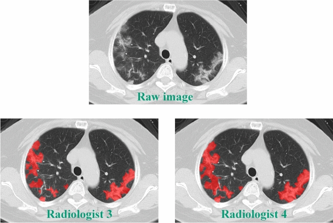
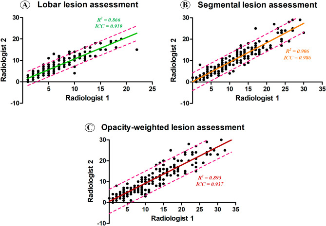
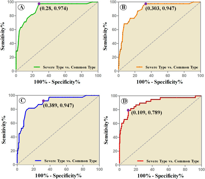
This study aimed to clarify and provide clinical evidence for which computed tomography (CT) assessment method can more appropriately reflect lung lesion burden of the COVID-19 pneumonia. A total of 244 COVID-19 patients were recruited from three local hospitals. All the patients were assigned to mild, common and severe types. Semi-quantitative assessment methods, e.g., lobar-, segmental-based CT scores and opacity-weighted score, and quantitative assessment method, i.e., lesion volume quantification, were applied to quantify the lung lesions. All four assessment methods had high inter-rater agreements. At the group level, the lesion load in severe type patients was consistently observed to be significantly higher than that in common type in the applications of four assessment methods (all the p < 0.001). In discriminating severe from common patients at the individual level, results for lobe-based, segment-based and opacity-weighted assessments had high true positives while the quantitative lesion volume had high true negatives. In conclusion, both semi-quantitative and quantitative methods have excellent repeatability in measuring inflammatory lesions, and can well distinguish between common type and severe type patients. Lobe-based CT score is fast, readily clinically available, and has a high sensitivity in identifying severe type patients. It is suggested to be a prioritized method for assessing the burden of lung lesions in COVID-19 patients.
Conflict of interest statement
The authors declare no competing interests.
Figures
References
Publication types
MeSH terms
Grants and funding
- 2017198897/Medical Health Science and Technology Project of Zhejiang Provincial Health Commission
- YG2020YQ17/the Biomedical Engineering Interdisciplinary Research Fund of Shanghai Jiao Tong University
- 2020XGZX036/Zhejiang University Special Scientific Research Fund for COVID-19 Prevention and Control
- 2017YFC0113400/National Key Research and Development Program of China
LinkOut - more resources
Full Text Sources
Other Literature Sources
Medical
