

The human EDAR 370V/A polymorphism affects tooth root morphology potentially through the modification of a reaction-diffusion system
- PMID: 33664401
- PMCID: PMC7933414
- DOI: 10.1038/s41598-021-84653-4
The human EDAR 370V/A polymorphism affects tooth root morphology potentially through the modification of a reaction-diffusion system
Abstract
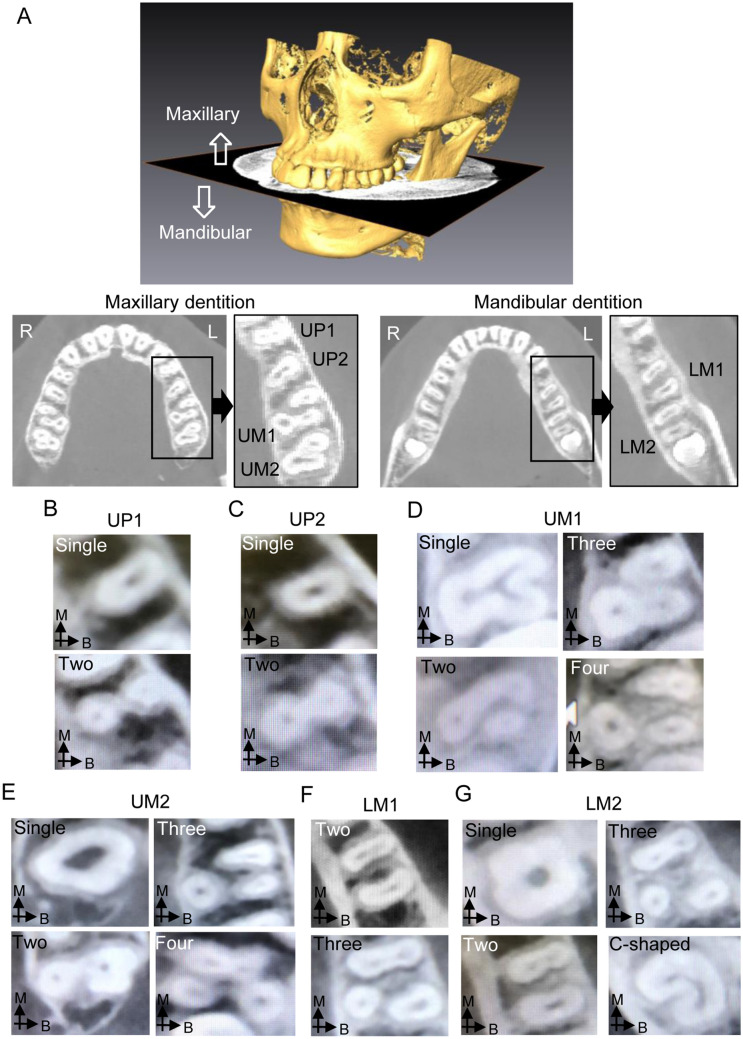
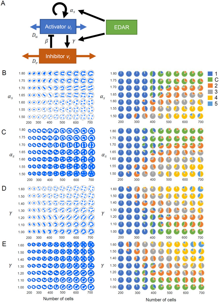
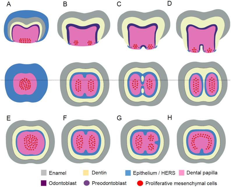
Morphological variations in human teeth have long been recognized and, in particular, the spatial and temporal distribution of two patterns of dental features in Asia, i.e., Sinodonty and Sundadonty, have contributed to our understanding of the human migration history. However, the molecular mechanisms underlying such dental variations have not yet been completely elucidated. Recent studies have clarified that a nonsynonymous variant in the ectodysplasin A receptor gene (EDAR 370V/A; rs3827760) contributes to crown traits related to Sinodonty. In this study, we examined the association between the EDAR polymorphism and tooth root traits by using computed tomography images and identified that the effects of the EDAR variant on the number and shape of roots differed depending on the tooth type. In addition, to better understand tooth root morphogenesis, a computational analysis for patterns of tooth roots was performed, assuming a reaction-diffusion system. The computational study suggested that the complicated effects of the EDAR polymorphism could be explained when it is considered that EDAR modifies the syntheses of multiple related molecules working in the reaction-diffusion dynamics. In this study, we shed light on the molecular mechanisms of tooth root morphogenesis, which are less understood in comparison to those of tooth crown morphogenesis.
Conflict of interest statement
The authors declare no competing interests.
Figures
Similar articles
-
Effects of an Asian-specific nonsynonymous EDAR variant on multiple dental traits.J Hum Genet. 2012 Aug;57(8):508-14. doi: 10.1038/jhg.2012.60. Epub 2012 May 31. J Hum Genet. 2012. PMID: 22648185
-
A common variation in EDAR is a genetic determinant of shovel-shaped incisors.Am J Hum Genet. 2009 Oct;85(4):528-35. doi: 10.1016/j.ajhg.2009.09.006. Am J Hum Genet. 2009. PMID: 19804850 Free PMC article.
-
Characterisation of a second gain of function EDAR variant, encoding EDAR380R, in East Asia.Eur J Hum Genet. 2020 Dec;28(12):1694-1702. doi: 10.1038/s41431-020-0660-6. Epub 2020 Jun 4. Eur J Hum Genet. 2020. PMID: 32499598 Free PMC article.
-
Ectodysplasin A (EDA) - EDA receptor signalling and its pharmacological modulation.Cytokine Growth Factor Rev. 2014 Apr;25(2):195-203. doi: 10.1016/j.cytogfr.2014.01.004. Epub 2014 Jan 23. Cytokine Growth Factor Rev. 2014. PMID: 24508088 Review.
-
Cellular and molecular mechanisms of tooth root development.Development. 2017 Feb 1;144(3):374-384. doi: 10.1242/dev.137216. Development. 2017. PMID: 28143844 Free PMC article. Review.
Cited by
-
Critical Considerations in Calling Disease-Causing EDAR Mutations in Nonsyndromic Oligodontia.J Clin Med. 2024 Dec 2;13(23):7328. doi: 10.3390/jcm13237328. J Clin Med. 2024. PMID: 39685785 Free PMC article.
-
EDA ligand triggers plasma membrane trafficking of its receptor EDAR via PKA activation and SNAP23-containing complexes.Cell Biosci. 2023 Jul 10;13(1):128. doi: 10.1186/s13578-023-01082-8. Cell Biosci. 2023. PMID: 37430358 Free PMC article.
-
Bioclimatic and masticatory influences on human cranial diversity verified by analysis of 3D morphometric homologous models.Sci Rep. 2024 Nov 4;14(1):26663. doi: 10.1038/s41598-024-76715-0. Sci Rep. 2024. PMID: 39496664 Free PMC article.
References
-
- Scott, G. R. & Turner, C. G. II. The Anthropology of Modern Human Teeth: Dental Morphology and Its Variation in Recent Human Populations (Cambridge University Press, 1997).
-
- Turner CG., II Advances in the dental search for native American origins. Acta Anthropogenet. 1984;8:23–78. - PubMed
Publication types
MeSH terms
Substances
LinkOut - more resources
Full Text Sources
Other Literature Sources
