

Telomeres and replicative cellular aging of the human placenta and chorioamniotic membranes
- PMID: 33664422
- PMCID: PMC7933277
- DOI: 10.1038/s41598-021-84728-2
Telomeres and replicative cellular aging of the human placenta and chorioamniotic membranes
Abstract
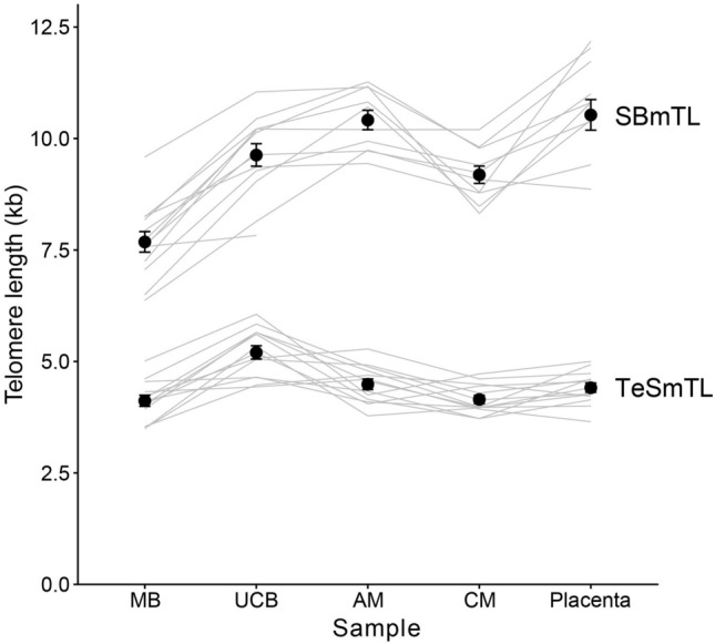
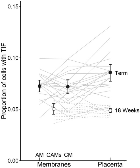
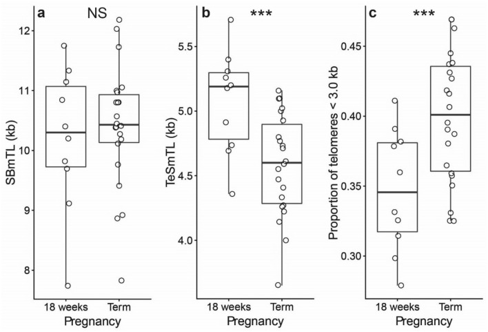
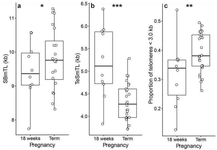
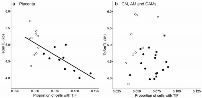
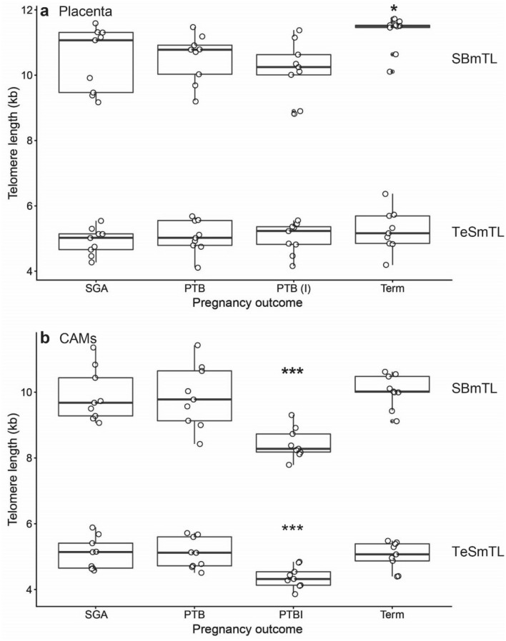
Recent hypotheses propose that the human placenta and chorioamniotic membranes (CAMs) experience telomere length (TL)-mediated senescence. These hypotheses are based on mean TL (mTL) measurements, but replicative senescence is triggered by short and dysfunctional telomeres, not mTL. We measured short telomeres by a vanguard method, the Telomere shortest length assay, and telomere-dysfunction-induced DNA damage foci (TIF) in placentas and CAMs between 18-week gestation and at full-term. Both the placenta and CAMs showed a buildup of short telomeres and TIFs, but not shortening of mTL from 18-weeks to full-term. In the placenta, TIFs correlated with short telomeres but not mTL. CAMs of preterm birth pregnancies with intra-amniotic infection showed shorter mTL and increased proportions of short telomeres. We conclude that the placenta and probably the CAMs undergo TL-mediated replicative aging. Further research is warranted whether TL-mediated replicative aging plays a role in all preterm births.
Conflict of interest statement
The authors declare no competing interests.
Figures
Similar articles
-
The telomere gestational clock: increasing short telomeres at term in the mouse.Am J Obstet Gynecol. 2019 May;220(5):496.e1-496.e8. doi: 10.1016/j.ajog.2019.01.218. Epub 2019 Jan 25. Am J Obstet Gynecol. 2019. PMID: 30690015
-
Telomere homeostasis in trophoblasts and in cord blood cells from pregnancies complicated with preeclampsia.Am J Obstet Gynecol. 2016 Feb;214(2):283.e1-283.e7. doi: 10.1016/j.ajog.2015.08.050. Epub 2015 Aug 28. Am J Obstet Gynecol. 2016. PMID: 26321036
-
[Replicative senescence as a model of aging: the role of oxidative stress and telomere shortening--an overview].Z Gerontol Geriatr. 1999 Apr;32(2):69-75. doi: 10.1007/s003910050086. Z Gerontol Geriatr. 1999. PMID: 10408009 Review. German.
-
Comparison of telomere length measurement methods.Philos Trans R Soc Lond B Biol Sci. 2018 Mar 5;373(1741):20160451. doi: 10.1098/rstb.2016.0451. Philos Trans R Soc Lond B Biol Sci. 2018. PMID: 29335378 Free PMC article. Review.
-
Does a sentinel or a subset of short telomeres determine replicative senescence?Mol Biol Cell. 2004 Aug;15(8):3709-18. doi: 10.1091/mbc.e04-03-0207. Epub 2004 Jun 4. Mol Biol Cell. 2004. PMID: 15181152 Free PMC article.
Cited by
-
Characterization of senescence-associated transcripts in the human placenta.Placenta. 2025 Mar 6;161:31-38. doi: 10.1016/j.placenta.2025.01.009. Epub 2025 Jan 20. Placenta. 2025. PMID: 39862734
-
Cellular senescence: from homeostasis to pathological implications and therapeutic strategies.Front Immunol. 2025 Feb 3;16:1534263. doi: 10.3389/fimmu.2025.1534263. eCollection 2025. Front Immunol. 2025. PMID: 39963130 Free PMC article. Review.
-
Buildup from birth onward of short telomeres in human hematopoietic cells.Aging Cell. 2023 Jun;22(6):e13844. doi: 10.1111/acel.13844. Epub 2023 Apr 28. Aging Cell. 2023. PMID: 37118904 Free PMC article.
-
Increase in short telomeres during the third trimester in human placenta.PLoS One. 2022 Jul 13;17(7):e0271415. doi: 10.1371/journal.pone.0271415. eCollection 2022. PLoS One. 2022. PMID: 35830448 Free PMC article.
-
The advancement of telomere quantification methods.Mol Biol Rep. 2021 Jul;48(7):5621-5627. doi: 10.1007/s11033-021-06496-6. Epub 2021 Jul 1. Mol Biol Rep. 2021. PMID: 34196896 Review.
References
Publication types
MeSH terms
Grants and funding
LinkOut - more resources
Full Text Sources
Other Literature Sources
