

Investigational Rho Kinase Inhibitors for the Treatment of Glaucoma
- PMID: 33664600
- PMCID: PMC7921633
- DOI: 10.2147/JEP.S259297
Investigational Rho Kinase Inhibitors for the Treatment of Glaucoma
Abstract
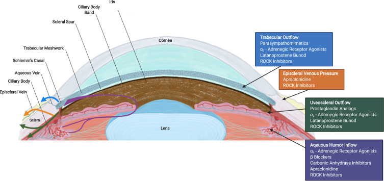
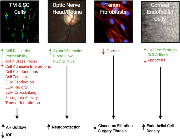
This review provides a comprehensive update on emerging ROCK inhibitors as an innovative treatment option for lowering intraocular pressure (IOP) in glaucoma and aims to describe the structure, mechanism of action, pharmaceutical characteristics, desirable ocular effects, including side effects for each agent. A literature review was conducted using PubMed, Scopus, clinicaltrials.gov, ARVO journals, Cochrane library and Selleckchem. Databases were searched using "investigational Rho kinase inhibitors," and "glaucoma" as keywords. In addition to this building block strategy, successive fractions were employed to further refine the results. Of the several ROCK inhibitors discovered, only two drugs are currently approved for glaucoma treatment; Netarsudil in the USA and Ripasudil in Japan and China. We identified and reviewed 15 agents currently in laboratory or clinical trials. These agents lower IOP mainly by decreasing outflow resistance through pharmacologic relaxation of the trabecular meshwork (TM) cells and reducing episcleral venous pressure. They have an optimistic safety profile; however, conjunctival hyperemia, conjunctival hemorrhage, pain on instillation, and corneal verticillata are common. Other properties such as neuroprotection (enhancing optic nerve blood flow and promoting axonal regeneration), anti-fibrotic activity, and endothelial cell proliferation may improve the visual prognosis and surgical outcomes in glaucoma. In addition, these agents have the potential to work synergistically with other topical glaucoma medications.
Keywords: ROCK; Schlemm’s canal; glaucoma; intraocular pressure; rho kinase inhibitors; trabecular meshwork.
© 2021 Al-Humimat et al.
Conflict of interest statement
The authors have no conflicts of interest in this work.
Figures
Similar articles
-
Rho-Kinase Inhibitors as Emerging Targets for Glaucoma Therapy.Ophthalmol Ther. 2023 Dec;12(6):2943-2957. doi: 10.1007/s40123-023-00820-y. Epub 2023 Oct 14. Ophthalmol Ther. 2023. PMID: 37837578 Free PMC article. Review.
-
Ripasudil hydrochloride hydrate: targeting Rho kinase in the treatment of glaucoma.Expert Opin Pharmacother. 2017 Oct;18(15):1669-1673. doi: 10.1080/14656566.2017.1378344. Epub 2017 Sep 14. Expert Opin Pharmacother. 2017. PMID: 28893104 Review.
-
Impact of the clinical use of ROCK inhibitor on the pathogenesis and treatment of glaucoma.Jpn J Ophthalmol. 2018 Mar;62(2):109-126. doi: 10.1007/s10384-018-0566-9. Epub 2018 Feb 14. Jpn J Ophthalmol. 2018. PMID: 29445943 Review.
-
Rho-kinase Inhibitors in Ocular Diseases: A Translational Research Journey.J Curr Glaucoma Pract. 2023 Jan-Mar;17(1):44-48. doi: 10.5005/jp-journals-10078-1396. J Curr Glaucoma Pract. 2023. PMID: 37228304 Free PMC article. Review.
-
Rho-kinase (ROCK) Inhibitors - A Neuroprotective Therapeutic Paradigm with a Focus on Ocular Utility.Curr Med Chem. 2020;27(14):2222-2256. doi: 10.2174/0929867325666181031102829. Curr Med Chem. 2020. PMID: 30378487 Review.
Cited by
-
The Effects of ROCK Inhibitor on Prevention of Dexamethasone-Induced Glaucoma Phenotype in Human Trabecular Meshwork Cells.Transl Vis Sci Technol. 2023 Dec 1;12(12):4. doi: 10.1167/tvst.12.12.4. Transl Vis Sci Technol. 2023. PMID: 38051267 Free PMC article.
-
Angiopoietin-1 Mimetic Nanoparticles for Restoring the Function of Endothelial Cells as Potential Therapeutic for Glaucoma.Pharmaceuticals (Basel). 2021 Dec 24;15(1):18. doi: 10.3390/ph15010018. Pharmaceuticals (Basel). 2021. PMID: 35056075 Free PMC article.
-
Role of Rho-Associated Protein Kinase Inhibition As Therapeutic Strategy for Parkinson's Disease: Dopaminergic Survival and Enhanced Mitophagy.Cureus. 2021 Aug 7;13(8):e16973. doi: 10.7759/cureus.16973. eCollection 2021 Aug. Cureus. 2021. PMID: 34377615 Free PMC article. Review.
-
Reticular epithelial corneal edema as a novel side-effect of Rho Kinase Inhibitors: An Indian scenario.Indian J Ophthalmol. 2022 Apr;70(4):1163-1170. doi: 10.4103/ijo.IJO_2865_21. Indian J Ophthalmol. 2022. PMID: 35326007 Free PMC article.
-
Rho Kinase (ROCK) Inhibitors in the Treatment of Glaucoma and Glaucoma Surgery: A Systematic Review of Early to Late Phase Clinical Trials.Pharmaceuticals (Basel). 2025 Apr 3;18(4):523. doi: 10.3390/ph18040523. Pharmaceuticals (Basel). 2025. PMID: 40283958 Free PMC article. Review.
References
Publication types
Grants and funding
LinkOut - more resources
Full Text Sources
Other Literature Sources
