Review
. 2020 Aug 29;8(1):4-10.
doi: 10.1093/nop/npaa055. eCollection 2021 Feb.

The cIMPACT-NOW updates and their significance to current neuro-oncology practice

Affiliations
Review

The cIMPACT-NOW updates and their significance to current neuro-oncology practice

L Nicolas Gonzalez Castro et al. Neurooncol Pract. .

Abstract

Over the past 4 years, advances in molecular pathology have enhanced our understanding of CNS tumors, providing new elements to refine their classification and improve the 2016 World Health Organization (WHO) Classification of CNS tumors. The Consortium to Inform Molecular and Practical Approaches to CNS Tumor Taxonomy-Not Official WHO (cIMPACT-NOW) was formed in late 2016 by a group of neuropathology and neuro-oncology experts to provide practical recommendations (published as cIMPACT-NOW updates) to improve the diagnosis and classification of CNS tumors, in advance of the publication of a new WHO Classification of CNS tumors. Here we review the content of all the available cIMPACT-NOW updates and discuss the implications of each update for the diagnosis and management of patients with CNS tumors.

Keywords: IDH-mutant gliomas; brain tumor classification; cIMPACT-NOW; glioblastoma; molecular pathology.


Figures

Figure 1.
Figure 1.
Example of oligodendroglioma, not elsewhere classified (NEC). In the hematoxylin-eosin–stained section, the histology of this tumor in the temporal lobe of a 40-year-old woman is fully compatible with a diffuse glioma (arrows indicate preexistent neurons that are overrun by diffusely infiltrating tumor cells). The tumor cells have a prototype oligodendroglial phenotype: “fried-egg” appearance with round nuclei and a clear halo. However, unlike “canonical” oligodendrogliomas as defined in the World Health Organization 2016 classification, this tumor was isocitrate dehydrogenase (IDH) wild-type and did not show codeletion of chromosome arms 1p and 19q. After ruling out other tumors that may have an oligodendroglial phenotype (especially dysembryoplastic neuroepithelial tumor, [extraventricular] neurocytoma, clear cell ependymoma, and pilocytic astrocytoma) and demonstrating that this tumor did not have molecular features of IDH–wild-type glioblastoma (see Consortium to Inform Molecular and Practical Approaches to CNS Tumor Taxonomy—Not Official WHO update 3), the tumor was signed out as “oligodendroglioma, NEC.”
Figure 2.
Figure 2.
Immunohistochemical surrogate markers enabling discrimination of astrocytoma, isocitrate dehydrogenase (IDH)-mutant from oligodendroglioma, IDH mutant. Hematoxylin-eosin–stained section of a tumor in a 52-year-old man reveals a diffuse, histologically low-grade glioma (preexistent neuron indicated by arrow). Using immunohistochemistry, this tumor can readily be characterized as IDH-mutant (tumor cells positive for IDH1 R132H mutant protein). In addition, because the tumor cell nuclei are strongly and extensively positive for p53 but negative for ATRX, immunohistochemistry allows for designating this tumor as astrocytoma, IDH-mutant (and discarding the diagnosis oligodendroglioma, IDH-mutant and 1p/19q-codeleted). The tumor microvasculature (indicated by arrowheads) serves as an internal control for the immunohistochemical stains (negative for IDH R132H–mutant protein and p53, positive for ATRX).
Figure 3.
Figure 3.
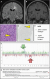
Histologically low-grade isocitrate dehydrogenase (IDH)–wild-type astrocytoma with molecular features of glioblastoma. A, T2–fluid-attenuated inversion recovery MRI of this 54-year-old male patient reveals a lesion that is compatible with diffuse low-grade glioma; in B, T1-weighed postgadolinium scans, the lesion is not enhancing. C and D, Hematoxylin-eosin staining of the needle biopsy material reveals only a slight increase in cellularity with mild nuclear pleomorphism and variably increased size of the (eosinophilic) cytoplasm, while Ki-67 staining reveals only very few proliferating cells (some positive nuclei indicated by arrowheads); these histopathological findings are compatible with the diagnosis of diffuse low-grade glioma. E, Next-generation sequencing of this lesion, however, reveals the absence of IDH1 and IDH2 mutation, but the presence of a TERT promoter mutation, meaning that the lesion according to cIMPACT-NOW update 3 qualifies as glioblastoma, IDH–wild-type, F, The fact that the copy number profile (obtained by performing methylome analysis) reveals gain of complete chromosome 7 combined with loss of complete chromosome 10 fully supports this diagnosis. G, Analysis of the methylome profile of this lesion using the Heidelberg Brain Tumor Classifier, also suggests the diagnosis glioblastoma, IDH–wild-type as best match; the low tumor cell content in this biopsy material may well explain the suboptimal score for this diagnosis (as well as the relatively subtle levels of chromosome 7 gain and chromosome 10 loss in F). cIMPACT-NOW, Consortium to Inform Molecular and Practical Approaches to CNS Tumor Taxonomy—Not Official WHO.
Figure 4.
Figure 4.
Homozygous cyclin-dependent kinase inhibitor 2A/B (CDKN2A/B) loss in isocitrate dehydrogenase (IDH)-mutant astrocytoma: grade 4. A and B, MRI images of a left frontal lobe lesion in a 45-year-old man demonstrating increased A, T2–fluid-attenuated inversion recovery signal and contrast enhancement on B, T1 postgadolinium sequences. C, Histology with infiltrating pleomorphic astrocytic cells with high nuclear-to-cytoplasmic ratios (arrowheads) and only a few scattered mitoses; because necrosis and microvascular proliferation were absent, the histopathological diagnosis was anaplastic astrocytoma, IDH-mutant, World Health Organization grade III (arrow indicates preexistent neuron). D, Fluorescent in situ hybridization assay to analyze CDKN2A copy number status. A CDKN2A locus-specific probe (red) and a chromosome 9 centromeric probe (CEP9; green) are hybridized and used to assess the CDKN2A copy number status scored in approximately 50 nuclei. A signal pattern of more than 10% of nuclei with no CDKN2A signal and at least one CEP9 signal is considered diagnostic of homozygous deletion of CDKN2A. The present sample demonstrated loss of CDKN2A signal and preservation of CEP9 signal in 24 of 50 cells counted, consistent with homozygous deletion of CDKN2A and thus reason to upgrade the tumor grade to IV.

Similar articles

Cited by

References

    1. Louis DN, Ohgaki H, Wiestler OD, et al. . World Health Organization Classification of Tumours of the Central Nervous System. 4th ed. Louis DN, Ohgaki H, Wiestler OD, Cavenee WK, eds. Lyon: International Agency for Research on Cancer; 2016.
    1. Louis DN, Perry A, Reifenberger G, et al. . The 2016 World Health Organization Classification of Tumors of the Central Nervous System: a summary. Acta Neuropathol. 2016;131(6):803–820. - PubMed
    1. Kristensen BW, Priesterbach-Ackley LP, Petersen JK, Wesseling P.. Molecular pathology of tumors of the central nervous system. Ann Oncol. 2019;30(8):1265–1278. - PMC - PubMed
    1. Louis DN, Aldape K, Brat DJ, et al. . Announcing cIMPACT-NOW: the Consortium to inform molecular and practical approaches to CNS tumor taxonomy. Acta Neuropathol. 2017;133(1):1–3. - PubMed
    1. Louis DN, Wesseling P, Paulus W, et al. . cIMPACT-NOW update 1: not otherwise specified (NOS) and not elsewhere classified (NEC). Acta Neuropathol. 2018;135(3):481–484. - PubMed