

Biomimetic strategies for tendon/ligament-to-bone interface regeneration
- PMID: 33665493
- PMCID: PMC7889437
- DOI: 10.1016/j.bioactmat.2021.01.022
Biomimetic strategies for tendon/ligament-to-bone interface regeneration
Abstract
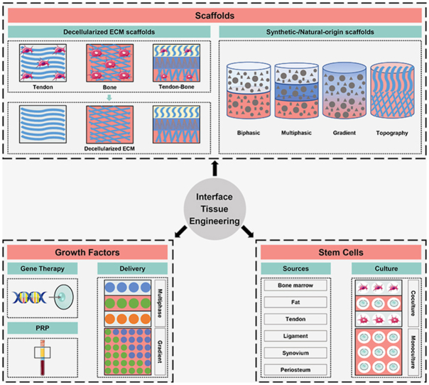
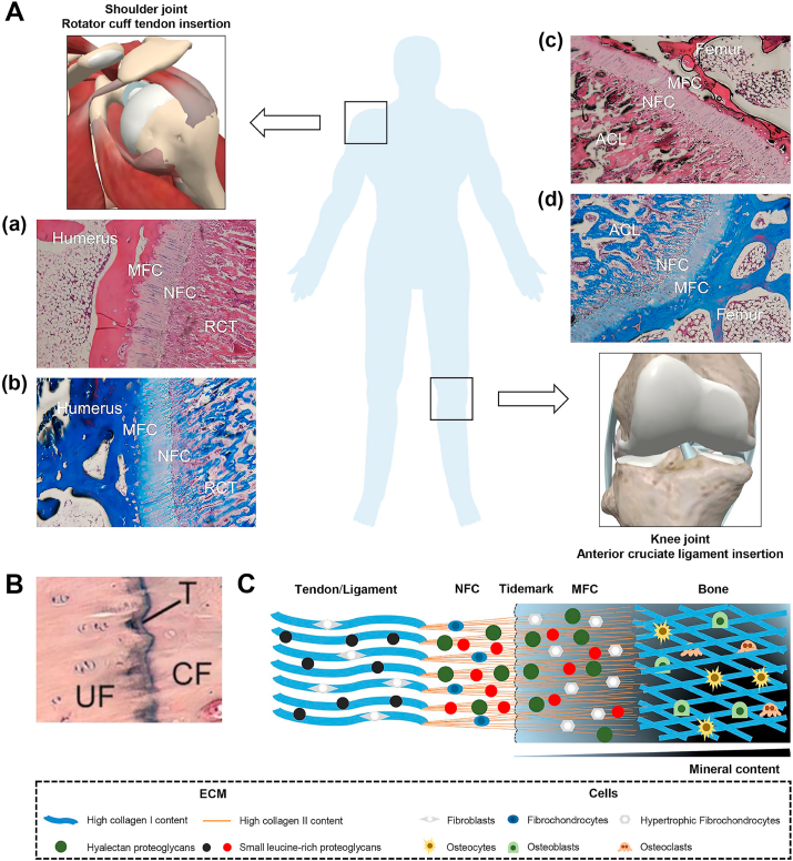
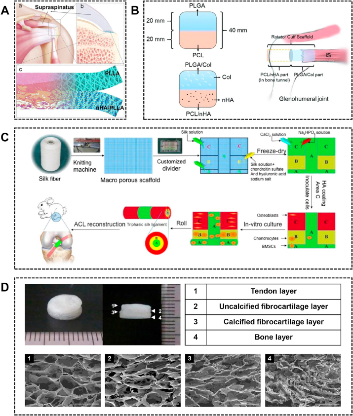
Tendon/ligament-to-bone healing poses a formidable clinical challenge due to the complex structure, composition, cell population and mechanics of the interface. With rapid advances in tissue engineering, a variety of strategies including advanced biomaterials, bioactive growth factors and multiple stem cell lineages have been developed to facilitate the healing of this tissue interface. Given the important role of structure-function relationship, the review begins with a brief description of enthesis structure and composition. Next, the biomimetic biomaterials including decellularized extracellular matrix scaffolds and synthetic-/natural-origin scaffolds are critically examined. Then, the key roles of the combination, concentration and location of various growth factors in biomimetic application are emphasized. After that, the various stem cell sources and culture systems are described. At last, we discuss unmet needs and existing challenges in the ideal strategies for tendon/ligament-to-bone regeneration and highlight emerging strategies in the field.
Keywords: Biomaterial; Growth factor; Stem cell; Tendon/ligament-to-bone interface; Tissue engineering.
© 2021 [The Author/The Authors].
Figures
Similar articles
-
[Application and prospect of tissue engineering strategies based on decellularized extracellular matrix in bone-tendon injuries].Zhonghua Yi Xue Za Zhi. 2023 Mar 21;103(11):787-792. doi: 10.3760/cma.j.cn112137-20220915-01959. Zhonghua Yi Xue Za Zhi. 2023. PMID: 36925110 Review. Chinese.
-
Biomimetic tendon extracellular matrix composite gradient scaffold enhances ligament-to-bone junction reconstruction.Acta Biomater. 2017 Jul 1;56:129-140. doi: 10.1016/j.actbio.2017.05.027. Epub 2017 May 11. Acta Biomater. 2017. PMID: 28502669
-
Heparin functionalization increases retention of TGF-β2 and GDF5 on biphasic silk fibroin scaffolds for tendon/ligament-to-bone tissue engineering.Acta Biomater. 2018 May;72:150-166. doi: 10.1016/j.actbio.2018.03.017. Epub 2018 Mar 14. Acta Biomater. 2018. PMID: 29550439
-
Biomimetic Scaffolds for Tendon Tissue Regeneration.Biomimetics (Basel). 2023 Jun 9;8(2):246. doi: 10.3390/biomimetics8020246. Biomimetics (Basel). 2023. PMID: 37366841 Free PMC article. Review.
-
Advances focusing on the application of decellularization methods in tendon-bone healing.J Adv Res. 2025 Jan;67:361-372. doi: 10.1016/j.jare.2024.01.020. Epub 2024 Jan 17. J Adv Res. 2025. PMID: 38237768 Free PMC article. Review.
Cited by
-
Engineering a functional ACL reconstruction graft containing a triphasic enthesis-like structure in bone tunnel for the enhancement of graft-to-bone integration.J Orthop Translat. 2024 Mar 22;45:155-167. doi: 10.1016/j.jot.2024.01.004. eCollection 2024 Mar. J Orthop Translat. 2024. PMID: 38559900 Free PMC article.
-
Engineering soft-hard tissue interfaces in dental and craniofacial system by spatially controlled bioactivities.Bioact Mater. 2024 Nov 25;45:246-256. doi: 10.1016/j.bioactmat.2024.11.030. eCollection 2025 Mar. Bioact Mater. 2024. PMID: 39659726 Free PMC article. Review.
-
Advances and challenges in biomaterials for tendon and enthesis repair.Bioact Mater. 2025 Feb 20;47:531-545. doi: 10.1016/j.bioactmat.2025.01.001. eCollection 2025 May. Bioact Mater. 2025. PMID: 40062342 Free PMC article. Review.
-
Tissue-engineered mesenchymal stem cell constructs alleviate tendinopathy by suppressing vascularization.Bioact Mater. 2024 Jul 2;36:474-489. doi: 10.1016/j.bioactmat.2024.06.029. eCollection 2024 Jun. Bioact Mater. 2024. PMID: 39055350 Free PMC article.
-
Comparative effect of skeletal stem cells versus bone marrow mesenchymal stem cells on rotator cuff tendon-bone healing.J Orthop Translat. 2024 Jun 20;47:87-96. doi: 10.1016/j.jot.2024.05.005. eCollection 2024 Jul. J Orthop Translat. 2024. PMID: 39007033 Free PMC article.
References
-
- Calejo I., Costa-Almeida R., Gomes M.E. Cellular complexity at the interface: challenges in enthesis tissue engineering. Adv. Exp. Med. Biol. 2019;1144:71–90. - PubMed
-
- Lu H.H., Spalazzi J.P. Biomimetic stratified scaffold design for ligament-to-bone interface tissue engineering. Comb. Chem. High Throughput Screen. 2009;12:589–597. - PubMed
Publication types
LinkOut - more resources
Full Text Sources
Other Literature Sources
