. 2021 May 18;143(20):1973-1986.
doi: 10.1161/CIRCULATIONAHA.120.049952. Epub 2021 Mar 5.

Malonate Promotes Adult Cardiomyocyte Proliferation and Heart Regeneration

Affiliations

Malonate Promotes Adult Cardiomyocyte Proliferation and Heart Regeneration

Jiyoung Bae et al. Circulation. .

Abstract

Background: Neonatal mouse cardiomyocytes undergo a metabolic switch from glycolysis to oxidative phosphorylation, which results in a significant increase in reactive oxygen species production that induces DNA damage. These cellular changes contribute to cardiomyocyte cell cycle exit and loss of the capacity for cardiac regeneration. The mechanisms that regulate this metabolic switch and the increase in reactive oxygen species production have been relatively unexplored. Current evidence suggests that elevated reactive oxygen species production in ischemic tissues occurs as a result of accumulation of the mitochondrial metabolite succinate during ischemia via succinate dehydrogenase (SDH), and this succinate is rapidly oxidized at reperfusion. Mutations in SDH in familial cancer syndromes have been demonstrated to promote a metabolic shift into glycolytic metabolism, suggesting a potential role for SDH in regulating cellular metabolism. Whether succinate and SDH regulate cardiomyocyte cell cycle activity and the cardiac metabolic state remains unclear.

Methods: Here, we investigated the role of succinate and SDH inhibition in regulation of postnatal cardiomyocyte cell cycle activity and heart regeneration.

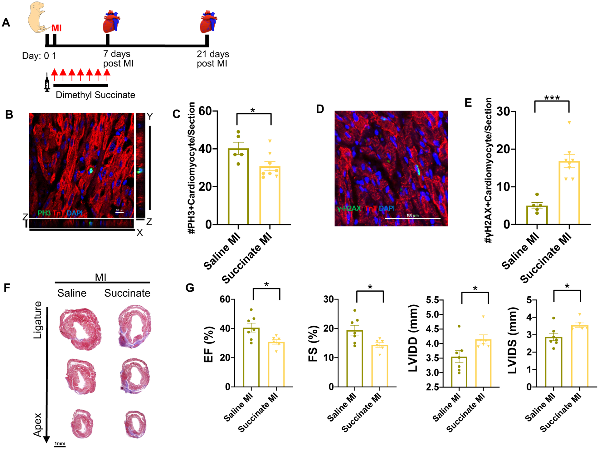
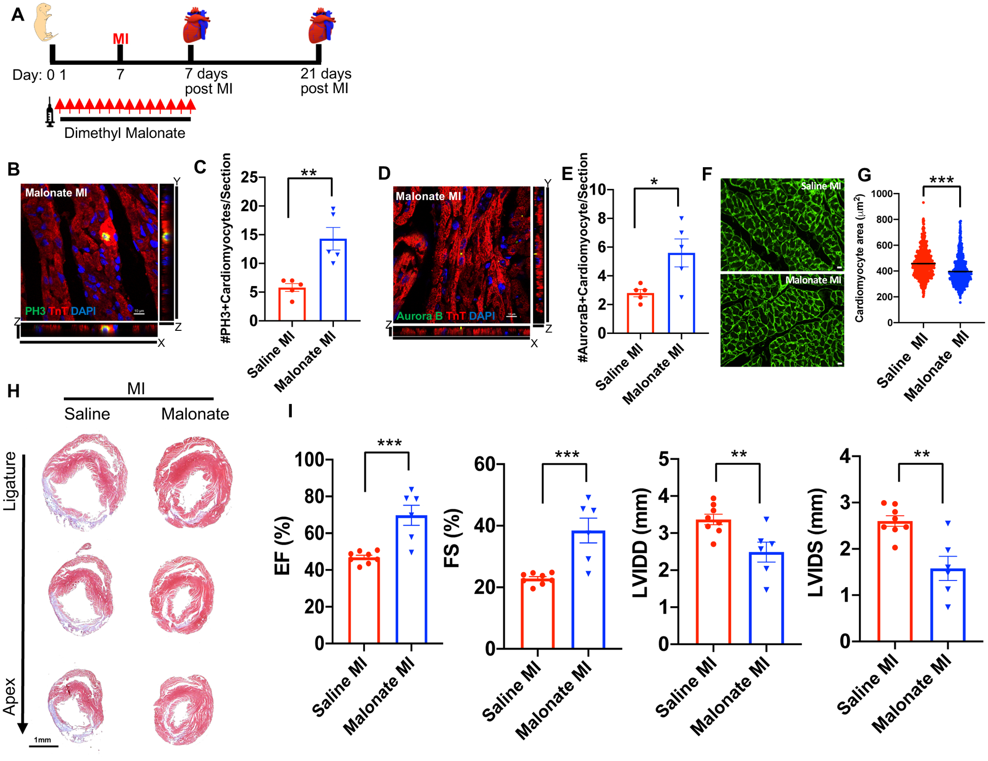
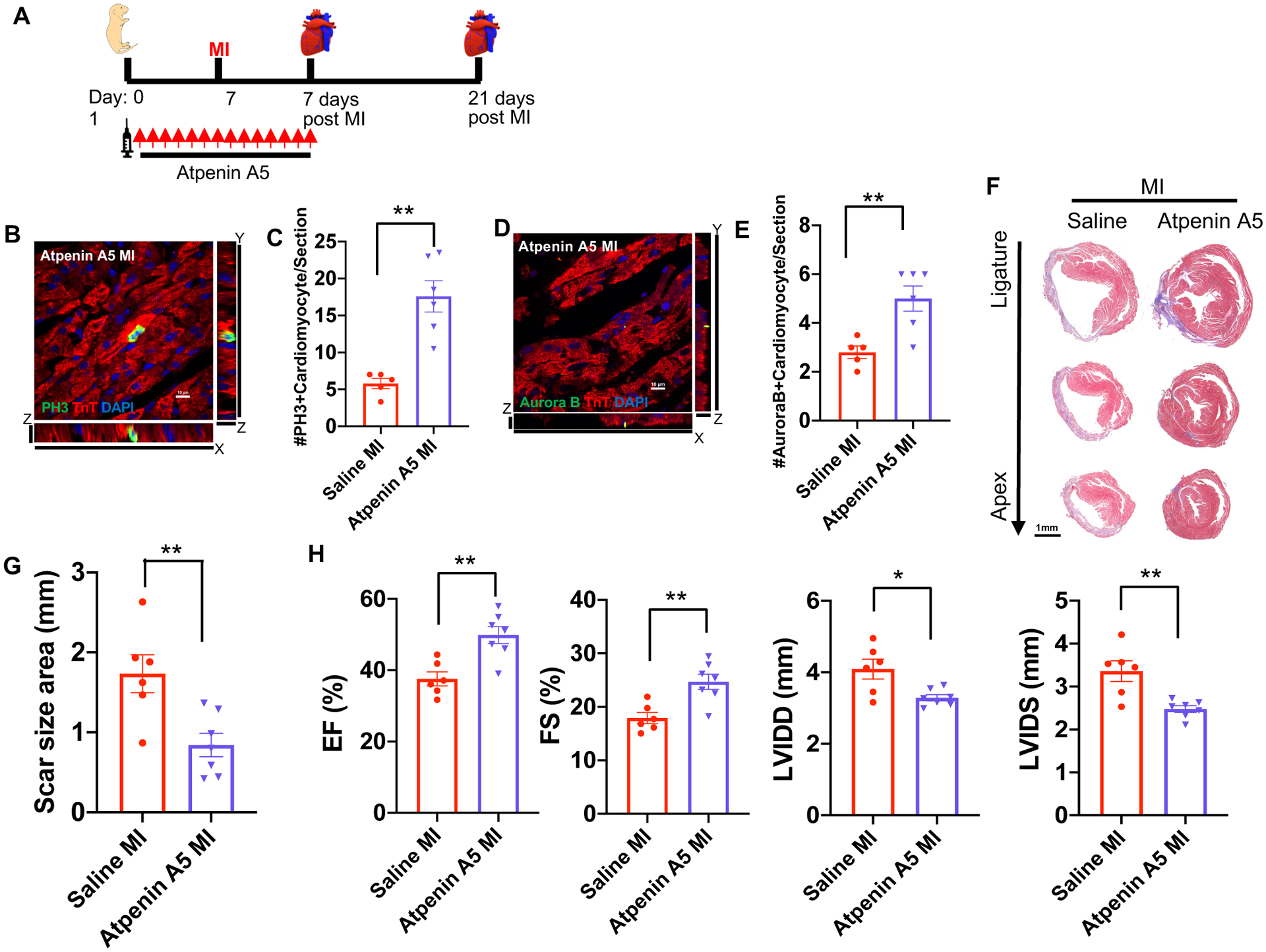
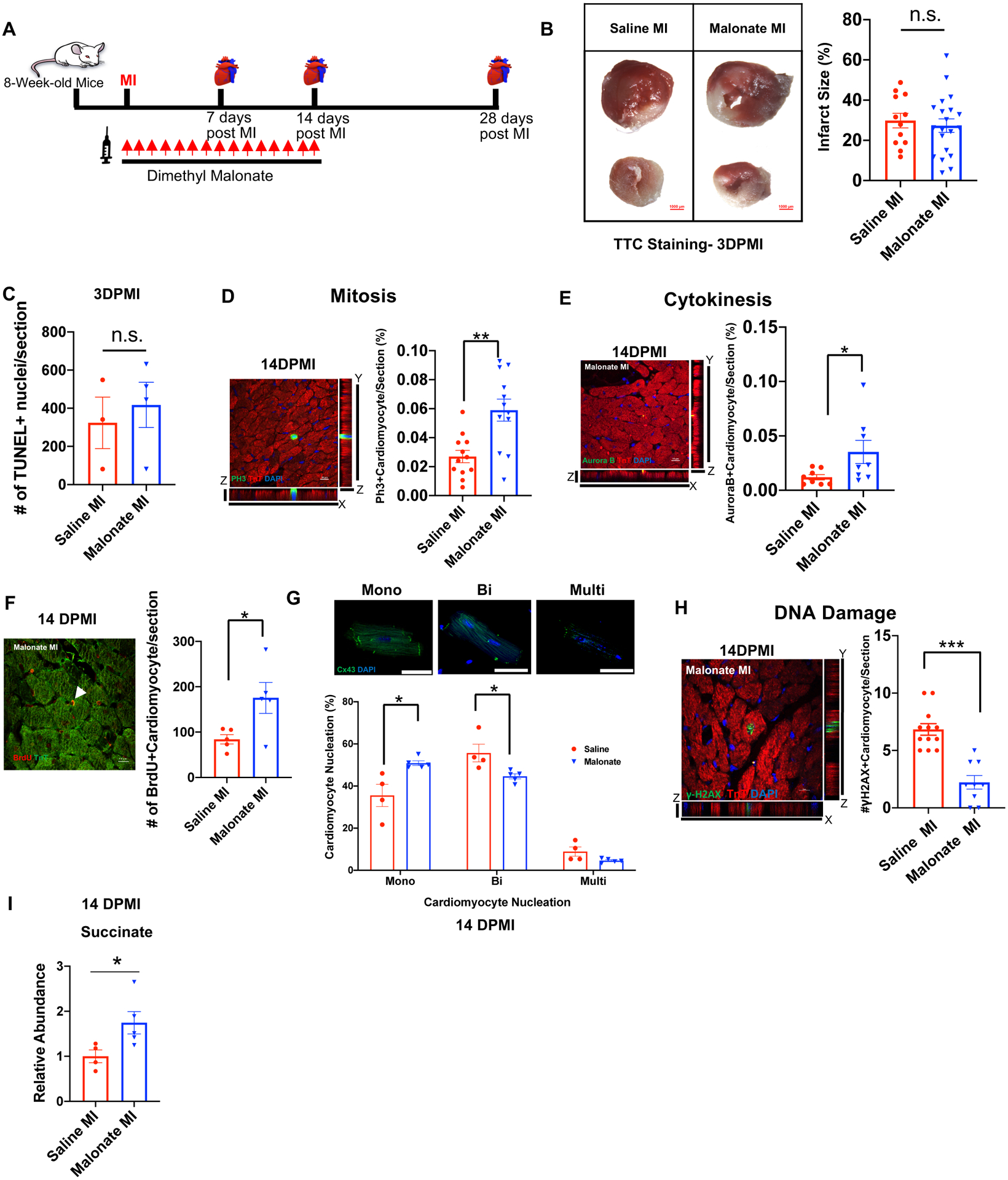
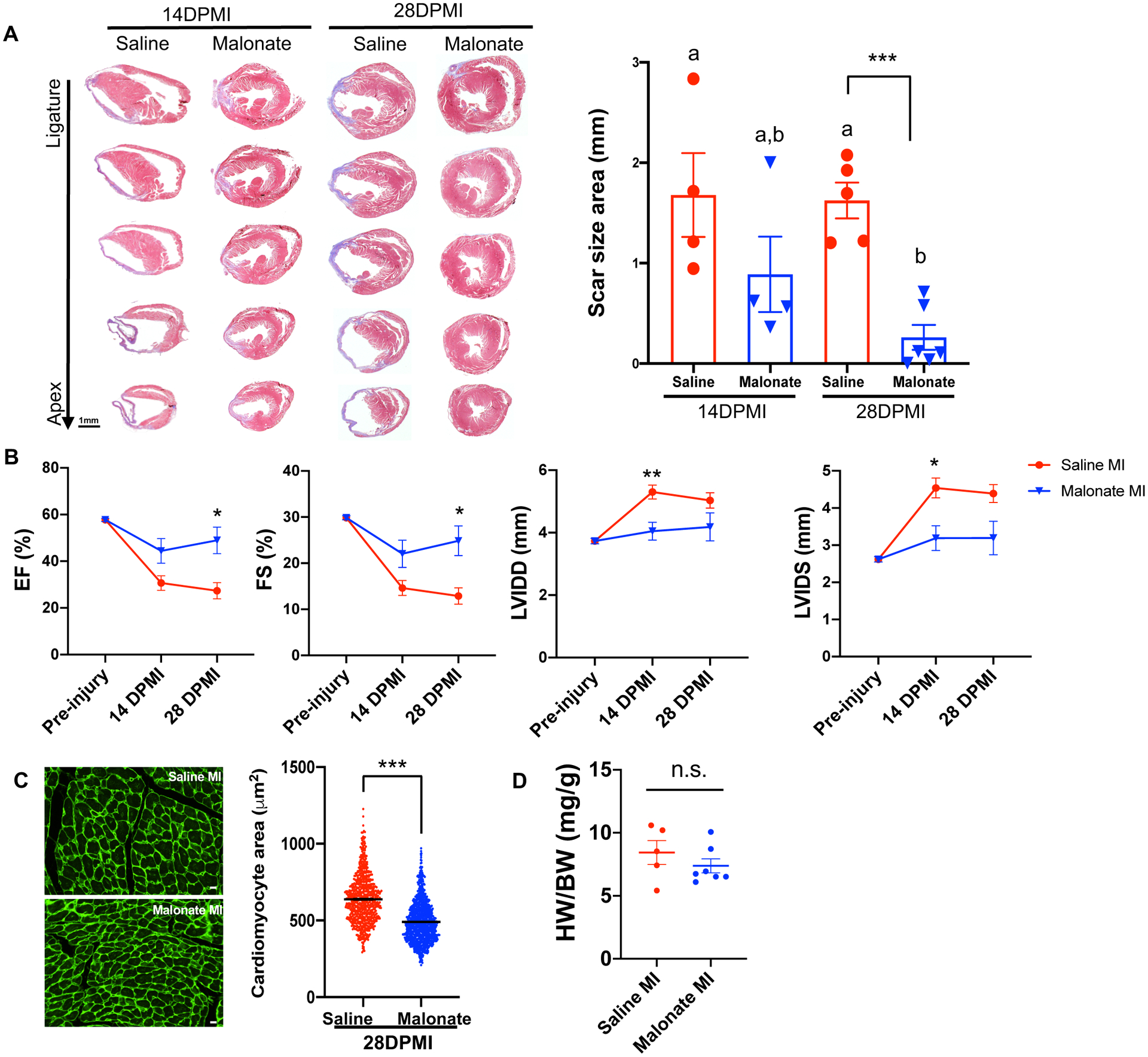
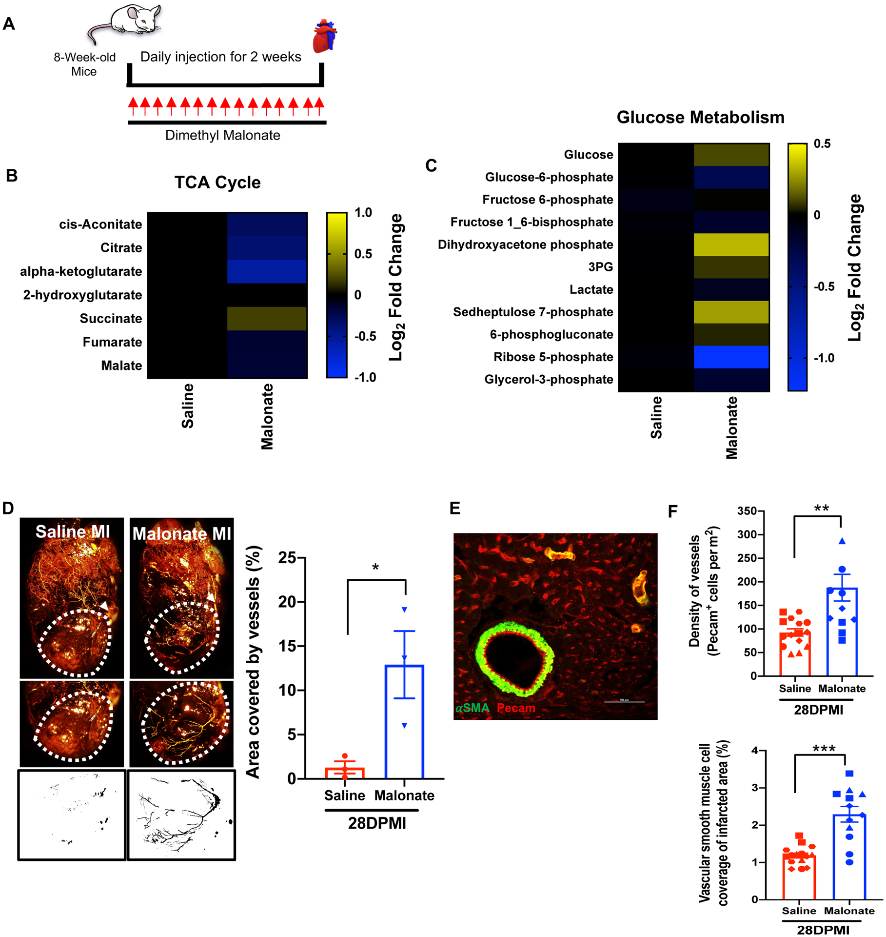
Results: Our results demonstrate that injection of succinate into neonatal mice results in inhibition of cardiomyocyte proliferation and regeneration. Our evidence also shows that inhibition of SDH by malonate treatment after birth extends the window of cardiomyocyte proliferation and regeneration in juvenile mice. Remarkably, extending malonate treatment to the adult mouse heart after myocardial infarction injury results in a robust regenerative response within 4 weeks after injury via promoting adult cardiomyocyte proliferation and revascularization. Our metabolite analysis after SDH inhibition by malonate induces dynamic changes in adult cardiac metabolism.

Conclusions: Inhibition of SDH by malonate promotes adult cardiomyocyte proliferation, revascularization, and heart regeneration via metabolic reprogramming. These findings support a potentially important new therapeutic approach for human heart failure.

Keywords: cell cycle; metabolism; myocardial infarction; regeneration; succinate dehydrogenase.


Conflict of interest statement

Disclosures

The authors declare no competing interests.

Figures

Figure 1.
Figure 1.. Succinate reduces cardiomyocyte proliferation and blocks heart regeneration in neonatal mice following myocardial infarction (MI).
(A) Schematic of injection period and myocardial infarction strategy in neonatal mice. (B) High magnification Z-stack image of a mitotic cardiomyocyte following immunostaining of pH3 and cTnT at 7 days post-MI. Scale bar, 10 μm. (C) Quantification of the number of mitotic cardiomyocytes per section showing a significant decrease in cardiomyocyte mitosis following dimethyl succinate injection. (D) Immunostaining of the DNA double-strand breaks marker γH2AX. Scale bar, 100 μm. (E) Quantification of cardiomyocytes with increased γH2AX foci demonstrating a significant increase in DNA damage in succinate-treated mice compared to controls. (F) Trichrome staining demonstrating persistence of the fibrotic scar following MI in the dimethyl succinate-injected mice compared to saline-injected controls. Scale bar, 1 mm. (G) Echocardiography at 21 days post-MI showing a significant reduction in the cardiac function of dimethyl succinate-injected mice following MI compared to saline-injected controls as measured by ejection fraction (EF), fractional shortening (FS), left ventricle internal diameter diastole (LVIDD) and left ventricle internal diameter systole (LVIDS). (n=5–8 mice per group). *P< 0.05, ***P<0.0001 by two-tailed unpaired Student’s t-test.
Figure 2.
Figure 2.. Malonate promotes cardiomyocyte proliferation and heart regeneration in the postnatal heart following MI.
(A) Schematic of dimethyl malonate injection and MI strategy. (B, C) Immunostaining and quantification of pH3 positive cardiomyocytes showing a significant increase in the levels of mitotic myocytes in dimethyl malonate injected hearts at 7 days post-MI compared to controls. Scale bar, 10 μm. (D, E) Immunostaining and quantification of Aurora B positive cardiomyocytes demonstrating a significant increase in myocyte cytokinesis in the dimethyl malonate injected mice. Scale bar, 10 μm. (F, G) Wheat germ agglutinin (WGA) staining and cell size quantification showing decrease in the cardiomyocyte size in malonate injected hearts at 21 days post-MI. Quantitative analyses represent counting of multiple fields from independent samples per group (~ 700 cells per group). Scale bar, 10 μm. (H) Trichrome staining of malonate injected hearts at 21 days post-MI at P7, showing complete regeneration in dimethyl malonate injected mice compared to control. Scale bar, 1 mm. (I) Left ventricular systolic function quantified by EF, FS, LVIDD and LVIDS at 3 weeks post-MI demonstrating functional recovery in dimethyl malonate injected MI hearts compared to the saline injected controls. (n=5–8 mice per group). *P< 0.05, **P<0.005, ***P<0.0001 by two-tailed unpaired Student’s t-test.
Figure 3.
Figure 3.. SDH inhibition by Atpenin A5 promotes cardiomyocyte mitosis and regeneration in the postnatal heart following MI.
(A) Schematic of Atpenin A5 injection and MI strategy. (B) Z-stack confocal image of a mitotic cardiomyocyte stained with pH3 and cTnT. Scale bar, 10 μm. (C) Quantification of mitotic cardiomyocytes showing a significant increase in the number of mitotic cardiomyocytes in Atpenin A5 injected mice at 7 days post-MI compared to controls. (D) Z-stack confocal image of an Aurora B positive cardiomyocyte. Scale bar, 10 μm. (E) Quantification of cardiomyocytes undergoing cytokinesis showing a significant increase in the number of cardiomyocytes in cytokinesis in Atpenin A5 injected mice at 7 days post-MI compared to controls. (F) Trichrome staining of Atpenin A5 injected mice at 3 weeks post-MI at P7 demonstrating myocardial regeneration and a significant reduction in scar size in Atpenin A5 injected mice compared to controls. Scale bar, 1 mm. (G) Quantification of scar size by ImageJ software from serial sections from ligature to apex. (H) Echocardiography measurements of EF, FS, LVIDD and LVIDS at 3 weeks post-MI showing restoration of cardiac function in Atpenin A5 injected mice compared to controls. (n=6–8 mice per group). *P< 0.05, **P<0.005 by two-tailed unpaired Student’s t-test.
Figure 4.
Figure 4.. Malonate promotes adult cardiomyocyte proliferation following MI.
(A) Schematic of dimethyl malonate injection following adult MI. (B) TTC viability stain and quantification showing no significant difference in myocardial necrosis (white) in both saline and dimethyl malonate injected mice at 3 days post-MI (DPMI). Scale bar, 1 mm. (C) Quantification of TUNEL positive cardiomyocytes demonstrating no significant difference between TUNEL positive myocytes in saline and malonate injected mice at 3 days post-MI. (D) Z-stack confocal image of a pH3 positive cardiomyocyte at 14-days post-MI. Scale bar, 10 μm. Quantification of the percentage of pH3 positive cardiomyocytes showing a significant increase in the numbers of mitotic cardiomyocytes in dimethyl malonate injected hearts at 14-days post-MI compared to controls. (E) Z-stack confocal image of an Aurora B positive cardiomyocyte at 14-days post-MI. Scale bar, 10 μm. Quantification of the percentage of Aurora B positive cardiomyocytes demonstrating a significant increase in the numbers of cardiomyocytes undergoing cytokinesis in dimethyl malonate injected hearts at 14-days post-MI compared to controls. (F) Representative image and quantification of BrdU positive cardiomyocytes demonstrating a significant increase in BrdU positive cardiomyocytes at 14 days post-MI in malonate-treated mice. Scale bar, 10 μm. (G) Cardiomyocyte nucleation staining with connexin 43 (Cx43) and DAPI and quantification at 14 days post-MI demonstrating a significant increase in mononucleated cardiomyocytes as well as a significant decrease in binucleated cardiomyocytes following malonate treatment. Scale bar, 50 μm. (H) Immunostaining and quantification of the DNA double-strand breaks marker γH2AX demonstrating a significant decrease in DNA damage in malonate-treated mice compared to controls. Scale bar, 10 μm. (I) Relative intracellular abundance of succinate showing a significant increase in succinate levels in malonate-treated mice at 14 days post-MI (n=3–6 mice per group). *P< 0.05, **P<0.005, ***P<0.0001 by two-tailed unpaired Student’s t-test. n.s. indicates not significant.
Figure 5.
Figure 5.. Malonate restores cardiac structure and function following adult MI.
(A) Trichrome staining of heart sections from saline and dimethyl malonate-injected mice at 14 and 28 days following adult MI, showing restoration of cardiac structure and no fibrotic scarring by 28 days post-MI in dimethyl malonate injected mice. Quantification of scar size demonstrating a significant reduction in fibrosis in dimethyl malonate treated mice at 28 days post-MI. (B) Echocardiography of cardiac function measured by EF, FS, LVIDD and LVIDS at 14- and 28-days post-MI showing a significant functional recovery in dimethyl malonate injected hearts compared to saline injected controls at 28 days post-MI. (C) Wheat germ agglutinin (WGA) staining and cell size quantification showing decrease in cardiomyocyte size in dimethyl malonate injected hearts at 4 weeks post-MI. Quantitative analyses represent counting of multiple fields from 5–6 independent samples per group (750 ~ 1000 cells per group). Scale bar, 10 μm. (D) Heart weight-to-body weight ratios at 28 days post-MI showing no significant difference between saline and malonate injected mice. (n=5–6 mice per group). *P< 0.05, **P< 0.005, ***P<0.0001 by two-tailed unpaired Student’s t-test. One-way ANOVA was performed by Tukey’s multiple comparison test to determine the differences of group mean among treatment groups. Different letters indicate significant differences among groups. n.s. indicates not significant.
Figure 6.
Figure 6.. Malonate induces a dynamic metabolic shift in the adult heart and promotes revascularization following MI.
(A) Schematic of malonate administration for metabolomics. (B & C) Metabolomic changes of tricarboxylic acid (TCA) cycle and glucose metabolism in saline and malonate treated mice at 14 days following treatment. Relative abundance of metabolites in malonate-treated mice is compared to saline-treated mice and presented as a heatmap on a log2 scale demonstrating a dynamic change in TCA cycle and glucose metabolism in malonate-treated mice. (D) Coronary vessel casting by MICROFIL injection at 28 days post-MI showing a significant increase in revascularization of the infarct zone in malonate-treated mice compared to controls. Quantification of vasculature in region of interest (ROI) by analyzing binarized images for grey level intensity by ImageJ demonstrating a significant increase in vascular density in the infarct zone. (E) Immunostaining with the endothelial marker PECAM and vascular smooth muscle cell marker α-SMA. Scale bar, 100 μm. (F) Quantification of vascular lineages demonstrating a significant increase in endothelial capillary density and vascular smooth muscle cells in the infarct zone at 28 days post-MI. (n=3–4 mice per group). *P< 0.05, **P<0.005, ***P<0.0001 by two-tailed unpaired Student’s t-test.
Figure 7.
Figure 7.. Malonate treatment starting 1-week post-MI promotes myocardial regeneration.
(A) Schematic of malonate injection and MI strategy. (B) Trichrome staining of heart sections from saline and malonate-injected mice at 6 weeks post-MI. (C) Scar size quantification by ImageJ from serial sections per heart showing a significant reduction in scar size in malonate treated mice at 6 weeks post-MI. (D) Serial echocardiography measurements of EF, FS, LVID, LVIDS showing significant functional recovery in malonate-injected mice over time compared to controls. (n=6 mice per group). *P< 0.05, **P<0.005, ***P<0.0001 by two-tailed unpaired Student’s t-test. Two-way ANOVA with Bonferroni’s post-hoc test was used to determine the difference among the treatment groups and different time points. #P<0.05.

Comment in

References

    1. Virani SS, Alonso A, Benjamin EJ, Bittencourt MS, Callaway CW, Carson AP, Chamberlain AM, Chang AR, Cheng S, Delling FN et al. Heart Disease and Stroke Statistics-2020 Update: A Report From the American Heart Association. Circulation. 2020;141:e139–e596. - PubMed
    1. Mahmoud AI, Porrello ER, Kimura W, Olson EN and Sadek HA. Surgical models for cardiac regeneration in neonatal mice. Nat Protoc. 2014;9:305–11. - PMC - PubMed
    1. Porrello ER, Mahmoud AI, Simpson E, Hill JA, Richardson JA, Olson EN and Sadek HA. Transient regenerative potential of the neonatal mouse heart. Science. 2011;331:1078–80. - PMC - PubMed
    1. Porrello ER, Mahmoud AI, Simpson E, Johnson BA, Grinsfelder D, Canseco D, Mammen PP, Rothermel BA, Olson EN and Sadek HA. Regulation of neonatal and adult mammalian heart regeneration by the miR-15 family. Proc Natl Acad Sci U S A. 2013;110:187–92. - PMC - PubMed
    1. Webster WS and Abela D. The effect of hypoxia in development. Birth Defects Res C Embryo Today. 2007;81:215–28. - PubMed

Publication types