. 2021 May;54(5):e13000.
doi: 10.1111/cpr.13000. Epub 2021 Mar 5.

RNA helicase DDX5 acts as a critical regulator for survival of neonatal mouse gonocytes

Affiliations

RNA helicase DDX5 acts as a critical regulator for survival of neonatal mouse gonocytes

Qing Xia et al. Cell Prolif. 2021 May.

Abstract

Objectives: Mammalian spermatogenesis is a biological process of male gamete formation. Gonocytes are the only precursors of spermatogonial stem cells (SSCs) which develop into mature spermatozoa. DDX5 is one of DEAD-box RNA helicases and expresses in male germ cells, suggesting that Ddx5 plays important functions during spermatogenesis. Here, we explore the functions of Ddx5 in regulating the specification of gonocytes.

Materials and methods: Germ cell-specific Ddx5 knockout (Ddx5-/- ) mice were generated. The morphology of testes and epididymides and fertility in both wild-type and Ddx5-/- mice were analysed. Single-cell RNA sequencing (scRNA-seq) was used to profile the transcriptome in testes from wild-type and Ddx5-/- mice at postnatal day (P) 2. Dysregulated genes were validated by single-cell qRT-PCR and immunofluorescent staining.

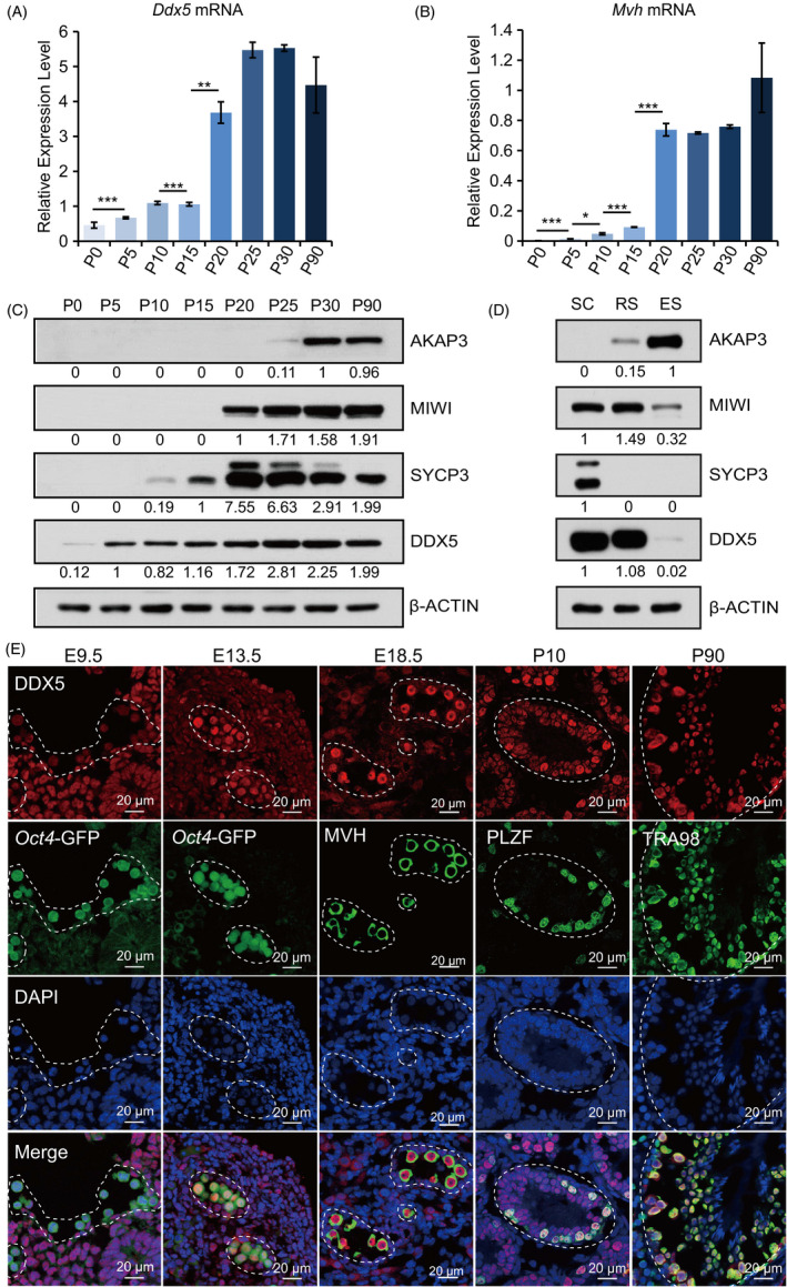
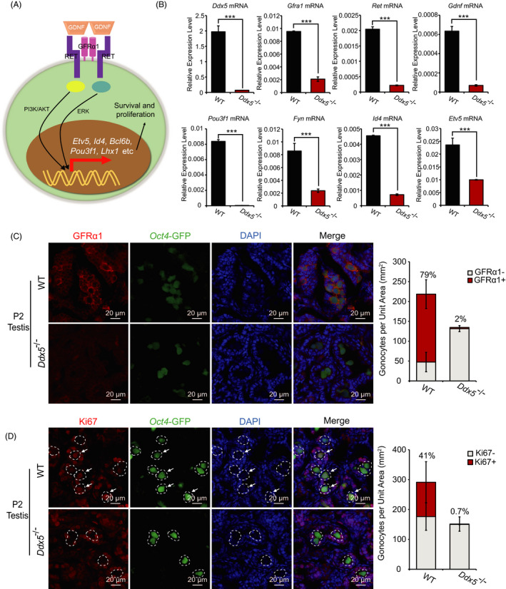
Results: In male mice, Ddx5 was expressed in germ cells at different stages of development. Germ cell-specific Ddx5 knockout adult male mice were sterile due to completely devoid of germ cells. Male germ cells gradually disappeared in Ddx5-/- mice from E18.5 to P6. Single-cell transcriptome analysis showed that genes involved in cell cycle and glial cell line-derived neurotrophic factor (GDNF) pathway were significantly decreased in Ddx5-deficient gonocytes. Notably, Ddx5 ablation impeded the proliferation of gonocytes.

Conclusions: Our study reveals the critical roles of Ddx5 in fate determination of gonocytes, offering a novel insight into the pathogenesis of male sterility.

Keywords: DDX5; RNA-binding protein; gonocyte; spermatogonial stem cell; testis.


Conflict of interest statement

The authors declare no competing interests.

Figures

FIGURE 1
FIGURE 1
Expression analysis of Ddx5 during development of male germline. A and B, qRT‐PCR to analyse the expression of Ddx5 and germ cell marker Mvh in the testes of mice at different stages (P0 to P90). Expression levels were normalized against geometric mean of Gapdh and Actin. Error bars correspond to means ± SD (*P < .05, **P < .01, ***P < .001). C, Western blotting to analyse DDX5 protein in the testes of mice at different stages (P0 to P90). AKAP3 was used as elongating spermatid marker. MIWI was used as spermatocyte and round spermatid marker. SYCP3 was used as spermatocytes marker. β‐ACTIN was used as a loading control. D, Western blotting to analyse protein level of AKAP3, MIWI, SYCP3 and DDX5 in the fractionated testicular spermatocytes (SC), round spermatids (RS) and elongating spermatids (ES). β‐ACTIN was used as a loading control. E, Immunofluorescent staining of DDX5 expression in germ cells from wild‐type mice at E9.5 to P90. Germ cells were labelled with Oct4‐GFP (green) in E9.5 and E13.5 gonad sections. Spermatogonia were labelled with anti‐PLZF antibody in testis section at P10. All germ cells except elongating spermatids were labelled with anti‐TRA98 antibody in testis section at P90. DNA was stained with DAPI (blue). Scale bars represent 20 μm
FIGURE 2
FIGURE 2
Ddx5 is essential for spermatogenesis. A, Schematic diagram of Ddx5 conditional knockout strategy. Left panel, the Ddx5 allele is flanked by two loxP sites. The blue squares stand for Ddx5 exon, and the red triangles represent loxP site. Right panel, reproductive strategy for Ddx5 ‐/‐ mice. B, Western blotting to detect protein level of DDX5, MIWI, MVH and SYCP3 in the testes of wild‐type (WT), Ddx5 +/‐ and Ddx5 ‐/‐ adult mice. β‐ACTIN was used as a loading control. C, Morphological analysis of testes in WT, Ddx5 +/‐ and Ddx5 ‐/‐ adult mice. Scale bar represents 5 mm. D, Assessment of testis to body weight ratio (mg/mg) from WT, Ddx5 +/‐ and Ddx5 ‐/‐ adult male mice. Error bars correspond to means ± SD. P = .4428 between WT and Ddx5 +/‐, P = 3.644e‐05 between Ddx5 +/‐ and Ddx5 ‐/‐ and P = 7.396e‐07 between WT and Ddx5 ‐/‐ (***P < .001, n = 6). E, H&E staining of testis sections from WT, Ddx5 +/‐ and Ddx5 ‐/‐ adult mice. The gaps between the seminiferous tubules due to the processing of testis section preparation. Scale bars represent 100 μm and 10 μm, respectively. SC, spermatocytes, ES, elongating spermatids, RS, round spermatids, SSC, spermatogonial stem cells, Sertoli, Sertoli cells. F, H&E staining of cauda epididymis sections from WT, Ddx5 +/‐ and Ddx5 ‐/‐ adult male mice. Scale bars represent 300 μm and 50 μm, respectively. G, Quantification of sperms released from cauda epididymides of WT, Ddx5 +/‐ and Ddx5 ‐/‐ adult males. Error bars correspond to means ± SD. P = .6646 between WT and Ddx5 +/‐, P = 1.027e‐05 between Ddx5 +/‐ and Ddx5 ‐/‐, and P = 1.467e‐10 between WT and Ddx5 ‐/‐ (***P < .001, n = 6)
FIGURE 3
FIGURE 3
Lack of germ cells in Ddx5 ‐/‐ testes. A, Immunofluorescent staining of DDX5 and TRA98 (germ cells) in testis sections from WT, Ddx5 +/‐ and Ddx5 ‐/‐ mice at P90. Null tubules were marked with asterisk. B, Immunofluorescent staining of DDX5 and SOX9 (Sertoli cells) in the testes from WT, Ddx5 +/‐ and Ddx5 ‐/‐ mice at P90. C, Immunofluorescent staining of DDX5 and TRA98 in testis sections from WT and Ddx5 ‐/‐ mice at P6. Null tubules were marked with asterisk. DNA was stained with DAPI. Scale bars represent 20 μm
FIGURE 4
FIGURE 4
Effects of Ddx5 knockout on the development of germ cells in neonatal mice. A, Oct4‐GFP expression in neonatal testes and ovaries in both WT and Ddx5 ‐/‐ mice. B, Immunofluorescent staining of DDX5 and TRA98 in the testes of WT, Ddx5 +/‐ and Ddx5 ‐/‐ mice at P0. Null tubules were marked with asterisk. DNA was stained with DAPI. Scale bars represent 20 μm. C, Numbers of TRA98‐positive germ cells (gonocytes) per unit area were counted in the testes of WT, Ddx5 +/‐ and Ddx5 ‐/‐ male mice at P0. Error bars correspond to means ± SD.P = .8534 between WT and Ddx5 +/‐, P = 1.083e‐05 between Ddx5 +/‐ and Ddx5 ‐/‐, and P = 1.083e‐05 between WT and Ddx5 ‐/‐ (***P < .001, n = 10). D, Oct4‐GFP‐positive gonocytes were analysed by FACS in testicular cells of WT, Ddx5 +/‐ and Ddx5 ‐/‐ mice at P2. FSC, forward scatter
FIGURE 5
FIGURE 5
Single‐cell RNA‐seq analysis of testes in both WT and Ddx5 ‐/‐ mice. A, Schematic overview of scRNA‐seq using neonatal mouse testis samples. B, Dimensionality reduction and clustering of testis scRNA‐seq data in WT and Ddx5 ‐/‐ mice at P2 (n = 11 278 cells). Colour coded for clustering analysis groups and annotated post hoc based on their transcriptional profile identities. Signature genes of clusters are listed in Table S3. C, UMAP plots of 5 major cell populations showed the expression of representatively well‐known cell type‐specific marker genes. Gene expression levels are indicated by shades of red. D, PCA plot based on the expression of highly variable genes (n = 3817) in WT (n = 47) and Ddx5 ‐/‐ (n = 7) germ cells. E, Volcano plot displaying DEGs between WT and Ddx5 ‐/‐ germ cells. Genes with a Benjamini‐Hochberg‐adjusted P value <.01 and an absolute value of log2 fold change (FC) >1 were considered significant. Red represented up‐regulated genes, green represented down‐regulated genes and grey represented non‐critical genes. The most significant DEGs in each direction were labelled. F, GO enrichment showing the terms associated with down‐regulated genes with adjusted P value lower than .01 from Ddx5 ‐/‐ germ cells. The dots indicated the GO categories with biological meaning. The colour of dots indicated high (red) or low (dark green) enrichment for a specific GO category. The size of dots displayed the overlap between the input gene lists with the collection of gene sets. G, Heat map showing the expression level of DEGs. The genes were ordered by annotated gene group and representative genes were shown. The colour gradient was row‐normalized across the samples based on log2 (TPM + 1)
FIGURE 6
FIGURE 6
Ddx5 maintains gonocytes and regulates the transcription of genes in GDNF pathway. A, Schematic diagram showing GDNF‐GFRα1‐RET signaling pathway in the gonocytes. B, Single‐cell qRT‐PCR to analyse the expression of Ddx5 and GDNF‐GFRα1‐RET signaling pathway‐related genes in Oct4‐GFP‐positive gonocytes sorted by FACS in WT and Ddx5 ‐/‐ testicular cells at P2. Expression levels were normalized against geometric mean of Gapdh and Actin. Error bars correspond to means ± SD (***P < .001, n = 3). C and D, Immunofluorescent staining of GFRα1 and Ki67 in WT and Ddx5 ‐/‐ testes at P2, respectively. Quantification of immunofluorescent signals of GFRα1 and Ki67 per unit area in the testis sections of WT and Ddx5 ‐/‐ male mice at P2, respectively, was calculated in the right panels. The percentage indicates the proportion of GFRα1 or Ki67‐positive gonocytes to all gonocytes in the testis sections. GFRα1‐: GFRα1‐negative gonocytes, GFRα1+: GFRα1‐positive gonocytes, Ki67‐: Ki67‐negative gonocytes, Ki67+: Ki67‐positive gonocytes. Error bars correspond to means ± SD (n = 3). Gonocytes were labelled with Oct4‐GFP (green). DNA was stained with DAPI. Scale bars represent 20 μm

Similar articles

Cited by

References

    1. Saitou M, Barton SC, Surani MA. A molecular programme for the specification of germ cell fate in mice. Nature. 2002;418:293‐300. - PubMed
    1. Saitou M. Specification of the germ cell lineage in mice. Front Biosci (Landmark Ed). 2009;14:1068‐1087. - PubMed
    1. Gunesdogan U, Magnusdottir E, Surani MA. Primordial germ cell specification: a context‐dependent cellular differentiation event [corrected]. Philos Trans R Soc Lond B Biol Sci. 2014;369:20130543. - PMC - PubMed
    1. Anderson R, Copeland TK, Scholer H, et al. The onset of germ cell migration in the mouse embryo. Mech Dev. 2000;91:61‐68. - PubMed
    1. Ginsburg M, Snow MH, McLaren A. Primordial germ cells in the mouse embryo during gastrulation. Development. 1990;110:521‐528. - PubMed

Substances