. 2021 Apr 1;27(19):6015-6027.
doi: 10.1002/chem.202005263. Epub 2021 Mar 5.

Azobioisosteres of Curcumin with Pronounced Activity against Amyloid Aggregation, Intracellular Oxidative Stress, and Neuroinflammation

Affiliations

Azobioisosteres of Curcumin with Pronounced Activity against Amyloid Aggregation, Intracellular Oxidative Stress, and Neuroinflammation

Julian Hofmann et al. Chemistry. .

Abstract

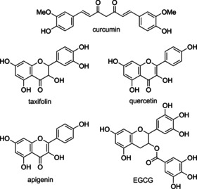
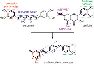
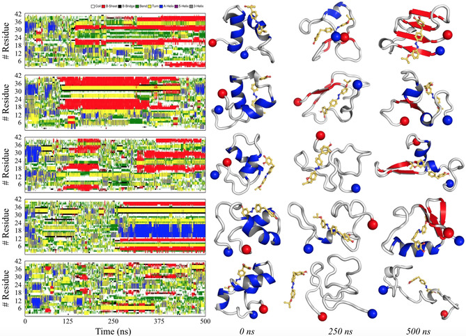
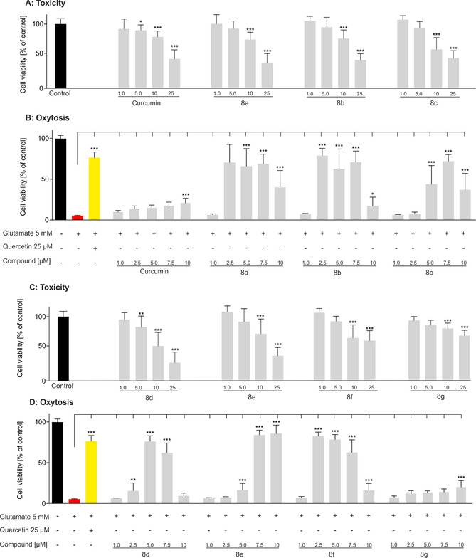
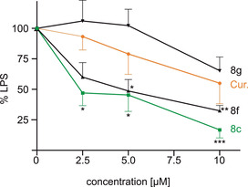
Many (poly-)phenolic natural products, for example, curcumin and taxifolin, have been studied for their activity against specific hallmarks of neurodegeneration, such as amyloid-β 42 (Aβ42) aggregation and neuroinflammation. Due to their drawbacks, arising from poor pharmacokinetics, rapid metabolism, and even instability in aqueous medium, the biological activity of azobenzene compounds carrying a pharmacophoric catechol group, which have been designed as bioisoteres of curcumin has been examined. Molecular simulations reveal the ability of these compounds to form a hydrophobic cluster with Aβ42, which adopts different folds, affecting the propensity to populate fibril-like conformations. Furthermore, the curcumin bioisosteres exceeded the parent compound in activity against Aβ42 aggregation inhibition, glutamate-induced intracellular oxidative stress in HT22 cells, and neuroinflammation in microglial BV-2 cells. The most active compound prevented apoptosis of HT22 cells at a concentration of 2.5 μm (83 % cell survival), whereas curcumin only showed very low protection at 10 μm (21 % cell survival).

Keywords: amyloid beta; bioisosterism; natural products; neuroprotectivity; replica-exchange molecular dynamics.


Conflict of interest statement

The authors declare no conflict of interest.

Figures

Figure 1
Figure 1
Chemical structures of curcumin, taxifolin, quercetin, apigenin, and epigallocatechin gallate (EGCG).
Figure 2
Figure 2
Rationalization for the azobioisostere prototype from the Aβ42 inhibitors curcumin and taxifolin. HBD=hydrogen‐bond donor; HBA=hydrogen‐bond acceptor.
Scheme 1
Scheme 1
Synthesis of target compounds 8 af and comparison compound 8 g. Reagents and conditions: i) FeCl3, 60 °C, 16 h; ii) 48 % HBr, AcOH, reflux, 3.5 h; iii) H2, Pd/C, MeOH, 10 bar, RT, 16 h; iv) oxone, CH2Cl2/H2O, RT, 3.5 h; v) AcOH, RT, 16 h, vi) nitrosobenzene, AcOH, RT, 16 h.
Figure 3
Figure 3
TEM analysis of the inhibitory effect on Aβ42. The Aβ monomer (100 μm) was incubated at 37 °C in phosphate‐buffered saline (PBS) for 24 h with or without 10 μm of the respective compound. A) Control; B) curcumin; C) 8 g; D) 8 f. Scale bar: 300 nm.
Figure 4
Figure 4
Left: Secondary‐structure analysis for the first five T‐replicas of Aβ42mon8 f, according to the DSSP algorithm. Right: Representative geometries for Aβ42mon8 f at 0, 250, and 500 ns of simulation. The N‐ and C‐terminal edges of Aβ42mon are reported as blue and red spheres, respectively.
Figure 5
Figure 5
Neuroprotection and neurotoxicity were determined in HT22 cells. 5 mm glutamate (red) induced cell death, 25 μm quercetin (yellow) served as a positive control for cell survival: A) neurotoxicity of curcumin and 8 ac; B) neuroprotection of curcumin and 8 ac; C) neurotoxicity of 8 dg; D) neuroprotection of 8 dg. Data are presented as means±SEM of three independent experiments and results refer to untreated control cells (black). Statistical analysis was performed by using one‐way analysis of variance (ANOVA) followed by Dunnett's multiple comparison post‐test by using GraphPad Prism 5, with reference to cells treated with 5 mm glutamate. Level of significance: ***p<0.001, **p<0.01, *p<0.05.
Figure 6
Figure 6
Effect of compounds 8 c, 8 f, 8 g, and curcumin on the production of NO as an inflammation marker. BV‐2 cells were treated with 50 ng mL−1 LPS alone or with the respective compound. NO was determined by the Griess assay in the supernatant. Data are presented as means±SEM of three independent experiments and results refer to LPS‐treated cells. Statistical analysis was performed by using one‐way ANOVA followed by Dunnett's multiple comparison post‐test by using GraphPad Prism 5. Level of significance: ***p<0.001, **p<0.01, *p<0.05.

Similar articles

Cited by

References

    1. E. Babusikova, A. Evinova, J. Jurecekova, M. Jesenak, D. Dobrota in Advanced Understanding of Neurodegenerative Diseases, IntechOpen, 2011.
    1. None
    1. Glenner G. G., Wong C. W., Biochem. Biophys. Res. Commun. 1984, 120, 885–890; - PubMed
    1. Masters C. L., Simms G., Weinman N. A., Multhaup G., McDonald B. L., Beyreuther K., Proc. Natl. Acad. Sci. USA 1985, 82, 4245–4249. - PMC - PubMed
    1. Haass C., Selkoe D. J., Nat. Rev. Mol. Cell Biol. 2007, 8, 101–112. - PubMed

LinkOut - more resources