. 2021 May 1;320(5):H1836-H1850.
doi: 10.1152/ajpheart.00881.2020. Epub 2021 Mar 5.

Fine particulate matter (PM2.5) inhalation-induced alterations in the plasma lipidome as promoters of vascular inflammation and insulin resistance

Affiliations

Fine particulate matter (PM2.5) inhalation-induced alterations in the plasma lipidome as promoters of vascular inflammation and insulin resistance

Bradford G Hill et al. Am J Physiol Heart Circ Physiol. .

Abstract

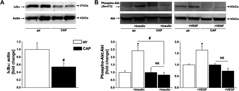
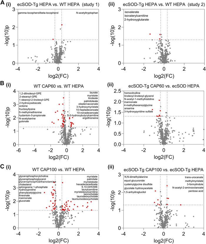
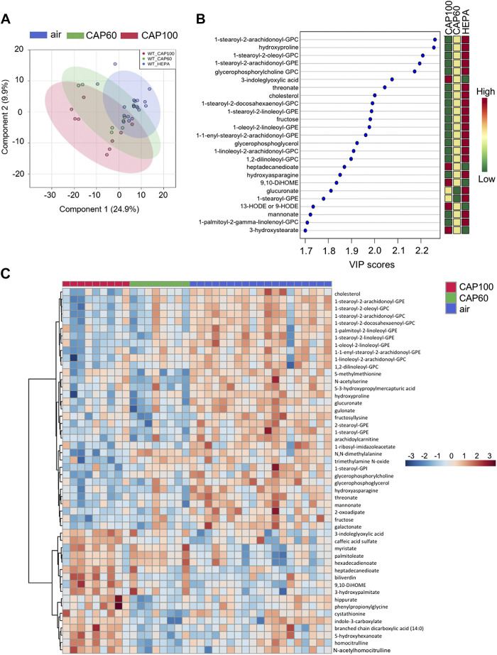
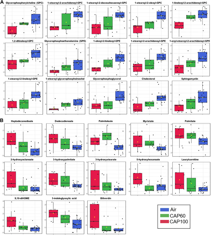
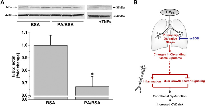
Fine particulate matter (PM2.5) air pollution exposure increases the risk of developing cardiovascular disease (CVD). Although the precise mechanisms by which air pollution exposure increases CVD risk remain uncertain, research indicates that PM2.5-induced endothelial dysfunction contributes to CVD risk. Previous studies demonstrate that concentrated ambient PM2.5 (CAP) exposure induces vascular inflammation and impairs insulin and vascular endothelial growth factor (VEGF) signaling dependent on pulmonary oxidative stress. To assess whether CAP exposure induces these vascular effects via plasmatic factors, we incubated aortas from naïve mice with plasma isolated from mice exposed to HEPA-filtered air or CAP (9 days) and examined vascular inflammation and insulin and VEGF signaling. We found that treatment of naïve aortas with plasma from CAP-exposed mice activates NF-κBα and induces insulin and VEGF resistance, indicating transmission by plasmatic factor(s). To identify putative factors, we exposed lung-specific ecSOD-transgenic (ecSOD-Tg) mice and wild-type (WT) littermates to CAP at concentrations of either ∼60 µg/m3 (CAP60) or ∼100 µg/m3 (CAP100) and measured the abundance of plasma metabolites by mass spectrometry. In WT mice, both CAP concentrations increased levels of fatty acids such as palmitate, myristate, and palmitoleate and decreased numerous phospholipid species; however, these CAP-induced changes in the plasma lipidome were prevented in ecSOD-Tg mice. Consistent with the literature, we found that fatty acids such as palmitate are sufficient to promote endothelial inflammation. Collectively, our findings suggest that PM2.5 exposure, by inducing pulmonary oxidative stress, promotes unique lipidomic changes characterized by high levels of circulating fatty acids, which are sufficient to trigger vascular pathology.NEW & NOTEWORTHY We found that circulating plasma constituents are responsible for air pollution-induced vascular pathologies. Inhalation of fine particulate matter (≤PM2.5) promotes a unique form of dyslipidemia that manifests in a manner dependent upon pulmonary oxidative stress. The air pollution-engendered dyslipidemic phenotype is characterized by elevated free fatty acid species and diminished phospholipid species, which could contribute to vascular inflammation and loss of insulin sensitivity.

Keywords: air pollution; cardiovascular disease; free fatty acids; plasma metabolome; pulmonary oxidative stress.


Conflict of interest statement

No conflicts of interest, financial or otherwise, are declared by the authors.

Figures

Figure 1.
Figure 1.
CAP exposure induces vascular injury via a plasmatic factor. Western blot analysis of inflammation (A) and insulin/VEGF resistance (B) in naïve aortas incubated with plasma isolated from mice exposed for 9 days to HEPA-filtered air or CAP. The aortas were treated with vehicle or stimulated with either insulin (100 nM) or VEGF (20 ng/mL) for 10 min. Western blot data are normalized to the vehicle or air controls. Data are means ± SE (*P < 0.05, insulin/VEGF vs. vehicle; #P < 0.05, air vs. CAP, n = 4). CAP, concentrated ambient PM2.5; NS, not significant; VEGF, vascular endothelial growth factor.
Figure 2.
Figure 2.
Overexpression of ecSOD in the lung prevents CAP-induced changes in the plasma metabolome. Volcano plots comparing plasma metabolite changes in ecSOD-Tg and WT mice under control conditions (HEPA-filtered air) and after CAP exposure: Mice were subjected to 9 days of exposure (6 h/day) to HEPA-filtered air, ∼60 µg/m3 CAP (CAP60, exposure 3), or ∼100 µg/m3 CAP (CAP100, exposure 4). Relative metabolite abundances in plasma were measured by unbiased metabolomic profiling. Metabolites that changed at least 1.25-fold with a raw P value <0.05 were considered statistically significant (indicated in red). The identities of some of the metabolites that changed significantly with CAP exposure are provided in the upper left and/or right quadrants of each volcano plot. A: plasma metabolite changes caused by ecSOD overexpression alone in metabolomics study I (i; n = 15 mice, 9 WT mice and 6 ecSOD-Tg mice) and metabolomics study II (ii; n = 19 mice, 10 WT mice and 9 ecSOD-Tg mice). B: plasma metabolite changes caused by CAP60 exposure in WT mice (i; n = 17 mice, 8 WT CAP60, 9 WT HEPA) and ecSOD-Tg mice (ii; n = 15 mice, 9 ecSOD-Tg CAP60 and 6 ecSOD-Tg HEPA). C: plasma metabolite changes caused by CAP100 exposure in WT mice (i; n = 19 mice, 9 WT CAP100 and 10 WT HEPA) and ecSOD-Tg mice (ii; n = 19 mice, 10 ecSOD-Tg CAP100 and 9 ecSOD-Tg HEPA). CAP, concentrated ambient PM2.5; ecSOD-Tg, extracellular superoxide dismutase-transgenic; WT, wild type.
Figure 3.
Figure 3.
Merged plasma metabolomic data reveal lipid species that are strongly influenced by CAP. Multivariate and heatmap metabolomic analyses of plasma from WT mice exposed to HEPA-filtered air, 60 µg/m3 CAP (CAP60) or 100 µg/m3 CAP (CAP100) for 9 days (6 h/day). Data from metabolomics study 1 (exposure 3) and metabolomics study 2 (exposure 4) were merged and each biochemical was rescaled to set the median equal to 1. Then, missing values were imputed with the minimum value for each biochemical. A: partial least squared discriminant analysis. B: variable importance in projection (VIP) analysis. C: heatmap analysis showing the 50 most significantly changed plasma metabolites in CAP-exposed mice (ANOVA). An FDR of <0.10 was considered statistically significant (n = 36 WT mice: 19 HEPA, 8 CAP60, and 9 CAP100). CAP, concentrated ambient PM2.5; FDR, false discovery rate; WT, wild type.
Figure 4.
Figure 4.
CAP concentration influences the circulating metabolome. Box plots of circulating metabolites that were significantly different in WT mice exposed to HEPA-filtered air, 60 µg/m3 CAP (CAP60) or 100 µg/m3 CAP (CAP100) for 9 days (6 h/day). Data from metabolomics study 1 (exposure 3) and metabolomics study 2 (exposure 4) were merged and each biochemical was rescaled to set the median equal to 1. Then, missing values were imputed with the minimum value for each biochemical. A: lipid species that decreased progressively with increasing CAP exposure. B: lipid species that increased progressively with increasing CAP exposure. ANOVA: an FDR of <0.10 was considered statistically significant (n = 36 WT mice: 19 HEPA, 8 CAP60, and 9 CAP100). CAP, concentrated ambient PM2.5; FDR, false discovery rate; WT, wild type.
Figure 5.
Figure 5.
Free fatty acids are sufficient to cause vascular pathology. Western blot analysis of IκBα in endothelial cells (A) incubated for 1 h with either bovine serum albumin (BSA, vehicle) or 100 µM palmitic acid (Sigma-Aldrich, complexed with BSA, PA/BSA). HUVEC incubated with TNF-α (10 ng/mL, 15 min) was used as a positive control. Western blot data are normalized to the vehicle controls. Data are means ± SE (*P < 0.05, PA/BSA vs. BSA, n = 3). B: inhalation of PM2.5 air pollution promotes a unique form of dyslipidemia that manifests in a manner dependent on pulmonary oxidative stress. This dyslipidemic phenotype is characterized by diminished phospholipid species and elevated free fatty acid species. Because elevated free fatty acids are sufficient to cause vascular inflammation and insulin or VEGF resistance, it is likely that PM2.5-induced dyslipidemia contributes to the increased CVD risk associated with air pollution. BSA, bovine serum albumin; HUVEC, human umbilical vein cell.

Comment in

References

    1. World Health Organization (WHO). https://www.who.int/health-topics/air-pollution#tab=tab_1.
    1. Brook RD, Rajagopalan S, Pope CA 3rd, Brook JR, Bhatnagar A, Diez-Roux AV, Holguin F, Hong Y, Luepker RV, Mittleman MA, Peters A, Siscovick D, Smith SC Jr, Whitsel L, Kaufman JD. Particulate matter air pollution and cardiovascular disease: an update to the scientific statement from the American Heart Association. Circulation 121: 2331–2378, 2010. doi:10.1161/CIR.0b013e3181dbece1. - DOI - PubMed
    1. Pope CA 3rd, Burnett RT, Thurston GD, Thun MJ, Calle EE, Krewski D, Godleski JJ. Cardiovascular mortality and long-term exposure to particulate air pollution: epidemiological evidence of general pathophysiological pathways of disease. Circulation 109: 71–77, 2004. - PubMed
    1. Pope CA 3rd, Turner MC, Burnett RT, Jerrett M, Gapstur SM, Diver WR, Krewski D, Brook RD. Relationships between fine particulate air pollution, cardiometabolic disorders, and cardiovascular mortality. Circ Res 116: 108–115, 2015. doi:10.1161/CIRCRESAHA.116.305060. - DOI - PubMed
    1. Pearson JF, Bachireddy C, Shyamprasad S, Goldfine AB, Brownstein JS. Association between fine particulate matter and diabetes prevalence in the U.S. Diabetes care 33: 2196–2201, 2010. doi:10.2337/dc10-0698. - DOI - PMC - PubMed

Publication types

MeSH terms

LinkOut - more resources