

Parafoveal Microvascular Alterations in Ocular and Non-Ocular Behҫet's Disease Evaluated With Optical Coherence Tomography Angiography
- PMID: 33666648
- PMCID: PMC7938019
- DOI: 10.1167/iovs.62.3.8
Parafoveal Microvascular Alterations in Ocular and Non-Ocular Behҫet's Disease Evaluated With Optical Coherence Tomography Angiography
Abstract
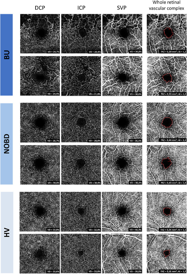
Purpose: To compare quantitative optical coherence tomography angiography (OCT-A) measurements of the parafoveal microvasculature in retinal capillary plexuses among Behҫet uveitis (BU) patients, non-ocular Behҫet's disease (NOBD) patients, and healthy volunteers (HVs).
Methods: Sixty-eight subjects were enrolled in this prospective observational cross-sectional study. OCT-A imaging was performed using the Heidelberg Engineering Spectralis OCT. A custom algorithm was developed to calculate the vessel density (VD) in three retinal vascular layers: deep capillary plexus, intermediate capillary plexus, and superficial vascular plexus. The foveal avascular zone (FAZ) and acircularity index were calculated for the whole retinal vascular complex.
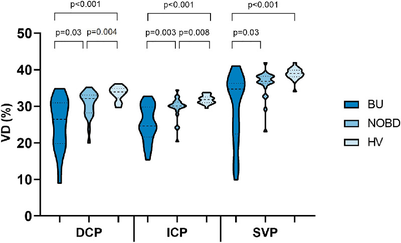
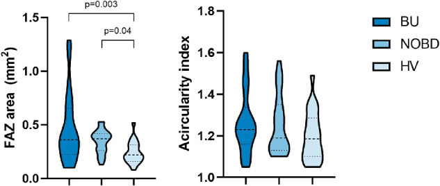
Results: We analyzed one eye from 21 BU patients (age, 51 ± 10 years), 23 NOBD patients (age, 48 ± 14 years), and 22 HVs (age, 44 ± 13 years). One-way multivariate analysis of covariance showed a statistically significant difference in VD among the three groups when combining the layers after controlling for scan quality (P < 0.001). The VD was lowest in the BU group and highest in the HV group in all layers. The FAZ area was also statistically significant different among the groups (P < 0.005), with the largest FAZ areas in BU patients and smallest FAZ areas in the HV group. However, no statistically significant difference was found for the acircularity index.
Conclusions: The parafoveal microvasculature is affected not only in BU patients but also in NOBD patients. Most deviations in the retinal microcirculation in Behҫet patients were found in the deeper layers of the retina by using the quantitative VD measurement.
Conflict of interest statement
Disclosure:
Figures
References
-
- Unoki N, Nishijima K, Kita M, Hayashi R, Yoshimura N. Structural changes of fovea during remission of Behçet's disease as imaged by spectral domain optical coherence tomography. Eye (Lond) . 2010; 24(6): 969–975. - PubMed
-
- Tugal-Tutkun I, Ozdal PC, Oray M, Onal S. Review for diagnostics of the year: multimodal imaging in Behçet uveitis. Ocul Immunol Inflamm . 2017; 25(1): 7–19. - PubMed
-
- Kappen J, van Dijk E, Baak-Dijkstra M, et al.. Behçet's disease, hospital-based prevalence and manifestations in the Rotterdam area. Neth J Med . 2015; 73(10): 471–477. - PubMed
-
- Kim M, Kim H, Kwon HJ, Kim SS, Koh HJ, Lee SC. Choroidal thickness in Behçet's uveitis: an enhanced depth imaging-optical coherence tomography and its association with angiographic changes. Invest Ophthalmol Vis Sci . 2013; 54(9): 6033–6039. - PubMed
-
- Cunningham ET Jr, Tugal-Tutkun I, Khairallah M, Okada AA, Bodaghi B, Zierhut M. Behçet uveitis. Ocul Immunol Inflamm . 2017; 25(1): 2–6. - PubMed
Publication types
MeSH terms
LinkOut - more resources
Full Text Sources
Other Literature Sources
Medical
