Review
. 2021 Mar 4;28(3):394-408.
doi: 10.1016/j.stem.2021.02.011.

Mitochondria as Signaling Organelles Control Mammalian Stem Cell Fate

Affiliations
Review

Mitochondria as Signaling Organelles Control Mammalian Stem Cell Fate

Ram Prosad Chakrabarty et al. Cell Stem Cell. .

Abstract

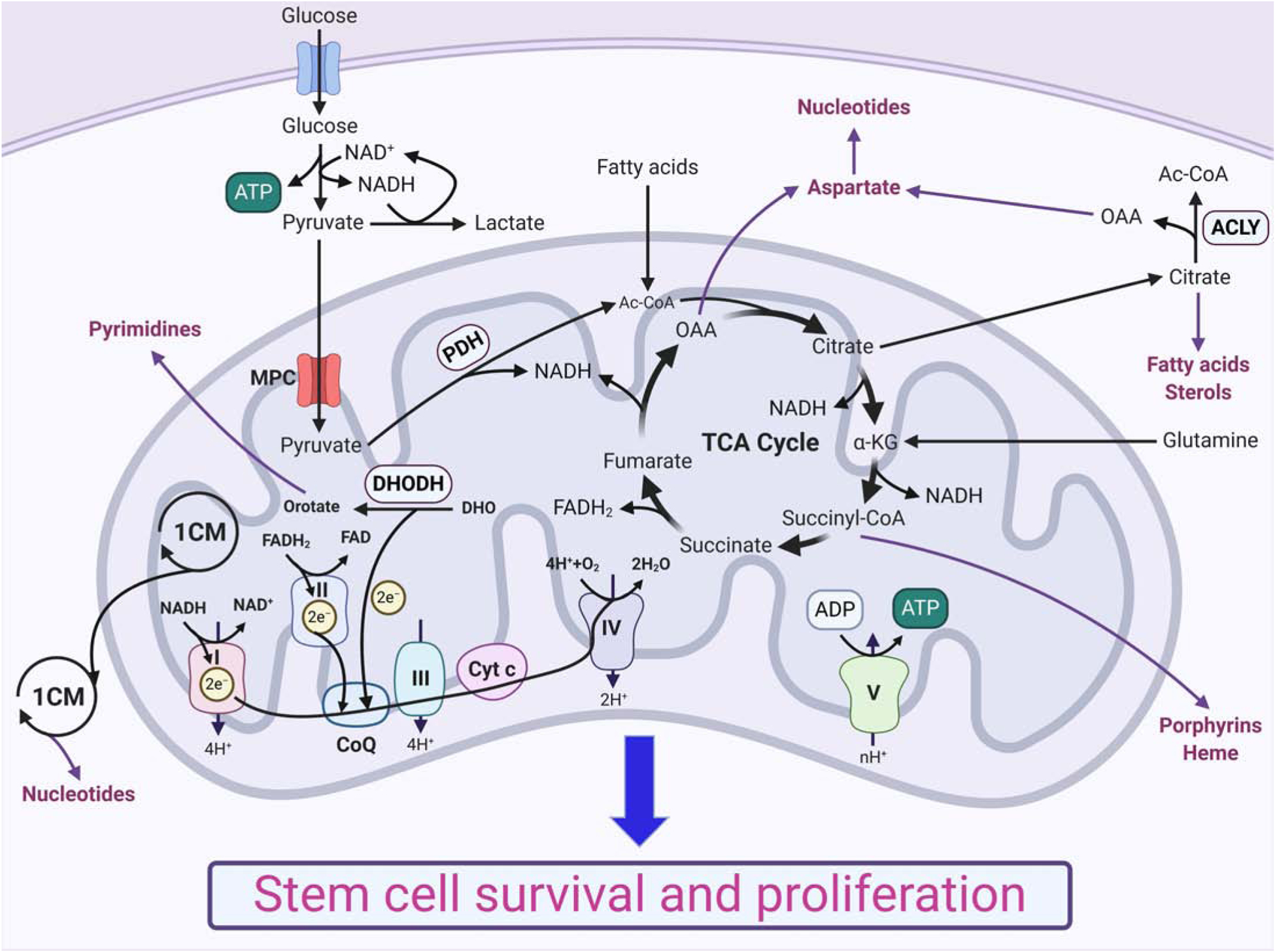
Recent evidence supports the notion that mitochondrial metabolism is necessary for the determination of stem cell fate. Historically, mitochondrial metabolism is linked to the production of ATP and tricarboxylic acid (TCA) cycle metabolites to support stem cell survival and growth, respectively. However, it is now clear that beyond these canonical roles, mitochondria as signaling organelles dictate stem cell fate and function. In this review, we focus on key conceptual ideas on how mitochondria control mammalian stem cell fate and function through reactive oxygen species (ROS) generation, TCA cycle metabolite production, NAD+/NADH ratio regulation, pyruvate metabolism, and mitochondrial dynamics.

Keywords: L-2-HG; ROS; TCA cycle; acetyl-CoA; epigenetics; mitochondrial dynamics; pyruvate.


Conflict of interest statement

Declaration of Interests The authors declare no competing interests.

Figures

Figure 1:
Figure 1:. Mitochondria as bioenergetic and biosynthetic organelles support stem cell survival and growth.
Glucose and multiple other substrates (e.g., fatty acids, glutamine) feed into the TCA cycle that generates biosynthetic intermediates and reducing equivalents, such as NADH and FADH2. Mitochondrial one-carbon metabolism (1CM) also provides biosynthetic intermediates. The intermediate substrates are used for the synthesis of macromolecules, such as lipids and nucleotides, and reducing equivalents fuel ATP generation by oxidative phosphorylation, which support stem cell survival and growth.
Figure 2:
Figure 2:. Mitochondria as signaling organelles regulate stem cell fate and function.
Mitochondria generate ROS and different metabolites that can act as cellular signals to control stem cell maintenance, commitment into progenitor populations, and differentiation into different cell types. (SC: Stem Cell; PC: Progenitor Cell; DC: Differentiated Cell)
Figure 3:
Figure 3:. Reactive oxygen species regulate stem cell fate and function.
(3a) Mitochondrial ETC and cytosolic NOX are two major sites of superoxide radical (O2) generation in a cell. Mitochondrial complex I generates O2 only in the mitochondrial matrix, whereas complex III generates O2 both in the matrix and intermembrane space. Immediately after generation, O2 gets converted into more stable and membrane-permeable hydrogen peroxide (H2O2) by superoxide dismutase (SOD2 in the matrix and SOD1 in the intermembrane space and cytosol). Mitochondrial O2 can also exit the mitochondria through voltage-dependent anion channels (VDAC) and get converted into H2O2 in the cytosol. H2O2 can oxidize specific sulfur-containing amino acids, primarily cysteine, of redox-sensitive proteins that are critical for stem cell fate and function and thereby control their functions, stability, and subcellular localization. Excess H2O2 can also generate hydroxyl radical (OH) through Fenton reaction, which is coupled with Fe2+ oxidation. OH can oxidize polyunsaturated fatty acids (PUFA) and generate lipid hydroperoxide that can induce cell death by ferroptosis. As excess ROS is toxic, cells have different ROS scavengers, such as peroxiredoxins (PRX) and glutathione peroxidases (GPX), as well, to maintain ROS homeostasis. (MOM: mitochondrial outer membrane; IM: intermembrane space; MIM: mitochondrial inner membrane) (3b) Stem cells require a low basal level of ROS for the maintenance of self-renewal capacity. FOXO proteins help maintain low ROS levels by activating the expression of different ROS scavengers. A moderate physiological increase in ROS level is necessary for commitment to progenitor cells and differentiation into different cell types. However, excess ROS accumulation leads to stem cell exhaustion. (SC: Stem Cell, PC: Progenitor Cell, DC: Differentiated Cell)
Figure 4:
Figure 4:. TCA cycle metabolites and NAD+/NADH ratio regulate stem cell fate and function through chromatin modifications.
Citrate derived acetyl-CoA is a substrate for histone acetyltransferase (HAT). Sirtuins, which are NAD+ dependent histone deacetylases, can remove histone acetyl marks. Mitochondria is a key regulator of the cellular NAD+/NADH ratio that regulates the activities of sirtuins. Similarly, mitochondrial one-carbon metabolism (1CM) contributes to the production of S-adenosyl methionine (SAM), which is a substrate for histone and DNA methyltransferases (KMTs, and DNMTs, respectively). Also, Jumonji C domain-containing ten-eleven translocation (TET) enzymes involved in the reversal of cytosine methylation and histone demethylases (KDMs) require α-KG, and these enzymes can be competitively inhibited by succinate, fumarate, and L-2-HG.
Figure 5:
Figure 5:. Pyruvate metabolism regulates stem cell fate and function.
Stem cells usually reduce glucose-derived pyruvate into lactate in the cytosol, whereas differentiated cells oxidize pyruvate through TCA cycle. In contrast to differentiated cells, stem cells have low levels of mitochondrial pyruvate carriers (MPC) that transports pyruvate from the cytosol into the mitochondria, and increased levels of uncoupling protein 2 (UCP2) that shunts away pyruvate from mitochondria into the cytosol, and pyruvate dehydrogenase kinases (PDKs) that inhibit pyruvate to acetyl-CoA conversion in the mitochondria. Stem cells also produce less H2O2 compared to their differentiated counterparts.

References

    1. Ahlqvist KJ, Hämäläinen RH, Yatsuga S, Uutela M, Terzioglu M, Götz A, Forsström S, Salven P, Angers-Loustau A, Kopra OH, et al. (2012). Somatic progenitor cell vulnerability to mitochondrial DNA mutagenesis underlies progeroid phenotypes in Polg mutator mice. Cell metabolism 15, 100–109. - PubMed
    1. Ansó E, Weinberg SE, Diebold LP, Thompson BJ, Malinge S, Schumacker PT, Liu X, Zhang Y, Shao Z, Steadman M, et al. (2017). The mitochondrial respiratory chain is essential for haematopoietic stem cell function. Nature cell biology 19, 614–625. - PMC - PubMed
    1. Avgustinova A, and Benitah SA (2016). Epigenetic control of adult stem cell function. Nature reviews. Molecular cell biology 17, 643–658. - PubMed
    1. Bahat A, and Gross A (2019). Mitochondrial plasticity in cell fate regulation. The Journal of biological chemistry 294, 13852–13863. - PMC - PubMed
    1. Baksh SC, and Finley LWS (2020). Metabolic Coordination of Cell Fate by α-Ketoglutarate-Dependent Dioxygenases. Trends in cell biology. - PMC - PubMed

Publication types

LinkOut - more resources