Review
. 2021 May 15:179:1-19.
doi: 10.1016/j.ijbiomac.2021.02.203. Epub 2021 Mar 2.

The human pandemic coronaviruses on the show: The spike glycoprotein as the main actor in the coronaviruses play

Affiliations
Review

The human pandemic coronaviruses on the show: The spike glycoprotein as the main actor in the coronaviruses play

Pedro F N Souza et al. Int J Biol Macromol. .

Abstract

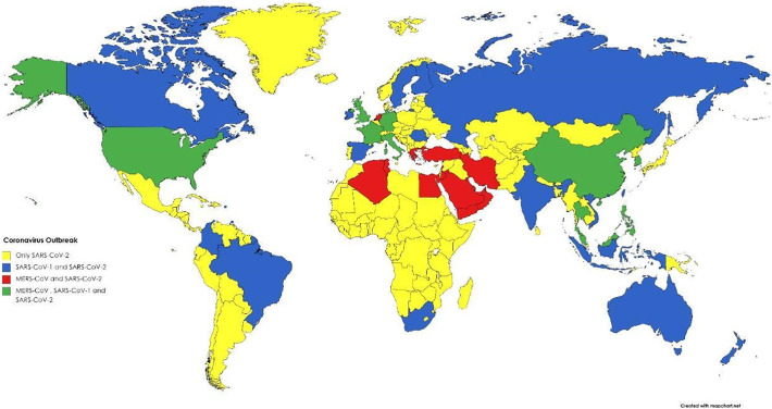
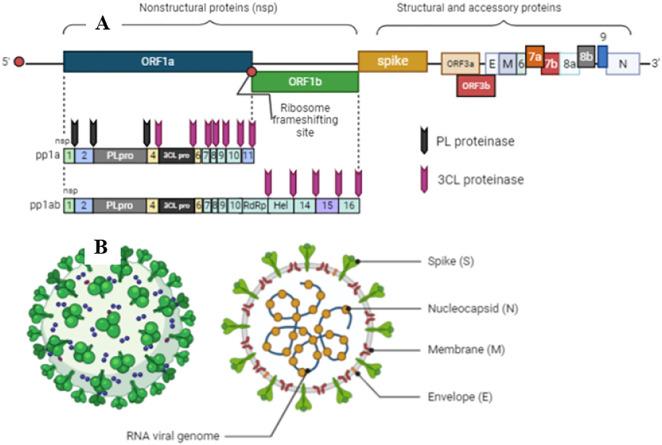
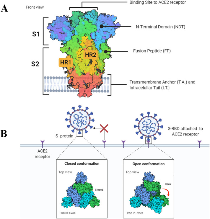
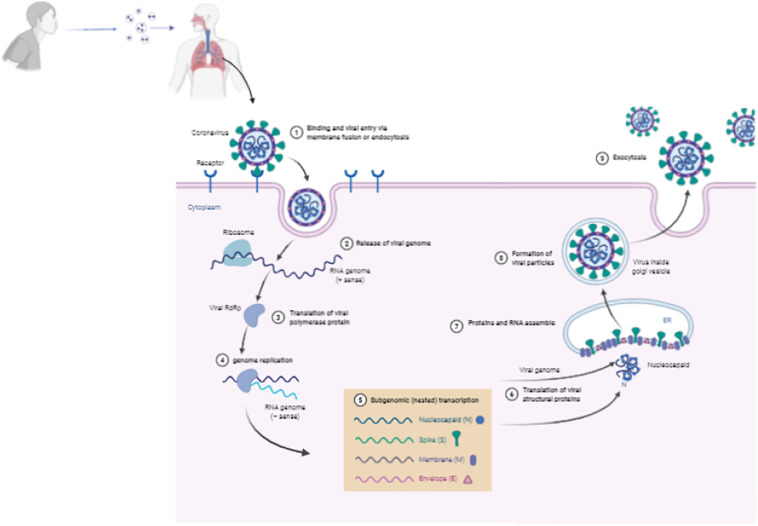
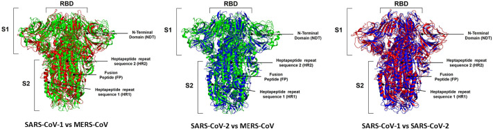
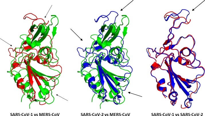
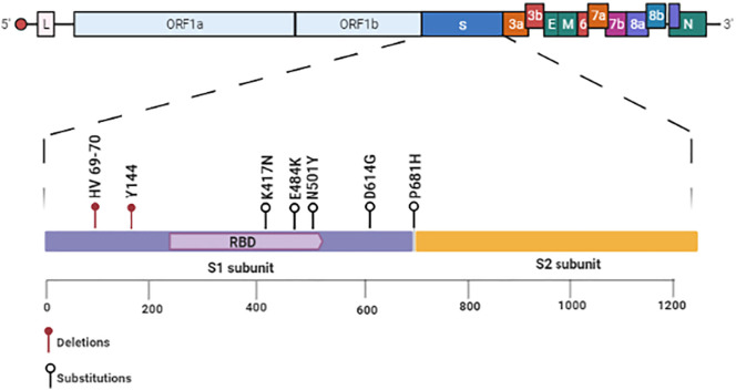
Three coronaviruses (CoVs) have threatened the world population by causing outbreaks in the last two decades. In late 2019, the severe acute respiratory syndrome coronavirus-2 (SARS-CoV-2) emerged and caused the coronaviruses to disease 2019 (COVID-19), leading to the ongoing global outbreak. The other pandemic coronaviruses, SARS-CoV and Middle East respiratory syndrome CoV (MERS-CoV), share a considerable level of similarities at genomic and protein levels. However, the differences between them lead to distinct behaviors. These differences result from the accumulation of mutations in the sequence and structure of spike (S) glycoprotein, which plays an essential role in coronavirus infection, pathogenicity, transmission, and evolution. In this review, we brought together many studies narrating a sequence of events and highlighting the differences among S proteins from SARS-CoV, MERS-CoV, and SARS-CoV-2. It was performed here, analysis of S protein sequences and structures from the three pandemic coronaviruses pointing out the mutations among them and what they come through. Additionally, we investigated the receptor-binding domain (RBD) from all S proteins explaining the mutation and biological importance of all of them. Finally, we discuss the mutation in the S protein from several new isolates of SARS-CoV-2, reporting their difference and importance. This review brings into detail how the variations in S protein that make SARS-CoV-2 more aggressive than its relatives coronaviruses and other differences between coronaviruses.

Keywords: Coronaviruses; MERS-CoV; Mutations; RBD; SARS-CoV; SARS-CoV-2; Spike proteins.


Conflict of interest statement

Declaration of competing interest All authors declare no conflicts of interest.

Figures

Fig. 1
Fig. 1
Worldwide view of Coronaviruses outbreak hotspots: spread locations of SARS-CoV 1, SARS-CoV 2, and MERS-CoV around the globe. Yellow represents dissemination of only SARS-CoV-2, blue represents dissemination of both SARS-CoV-1 and SARS-CoV-2, red represents dissemination of MERS-CoV and SARS-CoV-2, and green represents dissemination of the three coronaviruses. Created with BioRender.com.
Fig. 2
Fig. 2
Schematic diagram of coronaviruses genome and structural proteins of viral particles. A. Genome of coronaviruses produces non-structural proteins (nsp), such as RNA-dependent RNA polymerase involved in genome replication and two proteases involved in polyprotein processing, and structural and accessory proteins involved in composition of viral particles. B. The Genomes of coronaviruses also produce four structural proteins with significant roles in transmission and pathogenesis: spike (S), envelope (E), membrane (M), and nucleocapsid (N). Created with BioRender.com.
Fig. 3
Fig. 3
Schematic representation of the SARS-CoV-2 spike protein. A. Spike protein consists of the S1 and S2 units. In the S1 subunit, there is an extracellular N-terminal domain and a receptor-binding domain which play a role in the viral entrance into the cell through ACE2 receptor. In the S2 subunit, there is the fusion protein (FP), heptapeptide repeat sequence 1 and 2 (HR1 and HR2), as well as the transmembrane domain and a short C-terminal domain. B. Open or closed three dimensional conformation of S protein implicate in the binding of cell receptor ACE2. Created with BioRender.com.
Fig. 4
Fig. 4
Schematic model of coronavirus entry in cell. Coronaviruses bind to the host cell surface and release their RNA genomes into the cell through endocytosis. The positive-sense RNA genome is translated to produce the RNA-dependent RNA polymerase (RdRp) complex. Then the RdRp complex produces negative-sense RNA from the RNA genome, which provides the template for synthesis of positive-sense mRNAs. Then these subgenomic mRNAs are translated into structural and accessory proteins for viral particle assembly in endoplasmic reticulum. Finally, The enveloped virion is then exported from the cell by exocytosis. Created with BioRender.com.
Fig. 5
Fig. 5
Three-dimensional structure comparison of Spike protein among SARS-COV-1, MERS-CoV and SARS-COV2 through protein alignment. The spike protein of SARS-CoV-1 is represented in red, MERS-CoV in green and SARS-CoV-2 is blue. The 3D structures from S proteins MERS-CoV (PDB ID: 5X5C), SARS-CoV-1 (PDB ID: 5X58), and SARS-CoV-2 (PDB ID: 6Z97) deposited in the UniProt database deposited in the Protein Data Bank (PDB, https://www.rcsb.org/). RBD: Receptor-binding domain; NDT: N-terminal domain; HR1 and HR2: heptapeptide repeat sequence 1 and 2; FP: fusion peptide of subunit 1 and 2 are identified in the images. The structural alignments were performed in the Pymol program with the educational license.
Fig. 6
Fig. 6
Three-dimensional structure comparison of Receptor-Binding Domain (RBD) of Spike protein among SARS-COV-1, MERS-CoV and SARS-COV2 through protein alignment. The RBD of SARS-CoV-1 is represented in red, MERS-CoV in green and SARS-CoV-2 is blue. Structural differences are indicated by arrows. To perform the structural alignments was used the 3D structures of RBD from MERS-CoV (PDB ID: 6L8Q), SARS-CoV-1 (PDB ID: 2AJF), and SARS-CoV-2 (PDB ID: 6VW1) deposited in the UniProt database deposited in the Protein Data Bank (PDB, https://www.rcsb.org/). The structural alignments were performed in the Pymol program with the educational license.
Fig. 7
Fig. 7
Schematic diagram of mutations in the Spike protein from SARS-CoV-2. All relevant mutations in the Spike protein that lead to the new variants of SARS-CoV-2 are in the RBD. Among those, the most relevant are deletion: HV 69–70 and Y144. Substitutions: K417N, E484K, N501Y (RBD), D614G, and P681H. Created with BioRender.com.

References

    1. Weiss S.R. Forty years with coronaviruses. J. Exp. Med. 2020:217. - PMC - PubMed
    1. Coleman C.M., Frieman M.B. Coronaviruses: important emerging human pathogens. J. Virol. 2014;88:5209–5212. doi: 10.1128/jvi.03488-13. - DOI - PMC - PubMed
    1. Gorbalenya A.E., Baker S.C., Baric R.S., de Groot R.J., Drosten C., Gulyaeva A.A., Haagmans B.L., Lauber C., Leontovich A.M., Neuman B.W., et al. The species severe acute respiratory syndrome-related coronavirus: classifying 2019-nCoV and naming it SARS-CoV-2. Nat. Microbiol. 2020;5:536–544. - PMC - PubMed
    1. ICTV https://talk.ictvonline.org/taxonomy/ Available online: (accessed on Jan 21, 2021)
    1. Drosten C., Günther S., Preiser W., van der Werf S., Brodt H.-R., Becker S., Rabenau H., Panning M., Kolesnikova L., Fouchier R.A.M., et al. Identification of a novel coronavirus in patients with severe acute respiratory syndrome. N. Engl. J. Med. 2003;348:1967–1976. doi: 10.1056/nejmoa030747. - DOI - PubMed

MeSH terms

Substances