. 2021 Feb 13;7(2):136.
doi: 10.3390/jof7020136.

Direct Visualization of Fungal Burden in Filamentous Fungus-Infected Silkworms

Affiliations

Direct Visualization of Fungal Burden in Filamentous Fungus-Infected Silkworms

Yidong Yu et al. J Fungi (Basel). .

Abstract

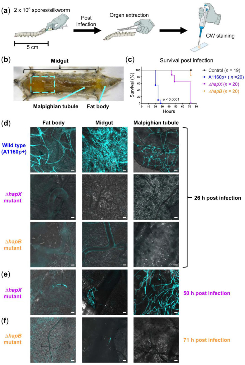
Invasive fungal infections (IFIs) are difficult to diagnose and to treat and, despite several available antifungal drugs, cause high mortality rates. In the past decades, the incidence of IFIs has continuously increased. More recently, SARS-CoV-2-associated lethal IFIs have been reported worldwide in critically ill patients. Combating IFIs requires a more profound understanding of fungal pathogenicity to facilitate the development of novel antifungal strategies. Animal models are indispensable for studying fungal infections and to develop new antifungals. However, using mammalian animal models faces various hurdles including ethical issues and high costs, which makes large-scale infection experiments extremely challenging. To overcome these limitations, we optimized an invertebrate model and introduced a simple calcofluor white (CW) staining protocol to macroscopically and microscopically monitor disease progression in silkworms (Bombyx mori) infected with the human pathogenic filamentous fungi Aspergillus fumigatus and Lichtheimia corymbifera. This advanced silkworm A. fumigatus infection model could validate knockout mutants with either attenuated, strongly attenuated or unchanged virulence. Finally, CW staining allowed us to efficiently visualize antifungal treatment outcomes in infected silkworms. Conclusively, we here present a powerful animal model combined with a straightforward staining protocol to expedite large-scale in vivo research of fungal pathogenicity and to investigate novel antifungal candidates.

Keywords: Aspergillus; Lichtheimia; calcofluor white staining; fungal infection model; silkworm.


Conflict of interest statement

The authors declare no conflict of interest.

Figures

Figure 1
Figure 1
Comparison of fungal burden in silkworms infected with different A. fumigatus strains. (a) Illustration of experimental setup; CW: Calcofluor white; (b) anatomy of silkworm: Dashed square indicates the area of midgut taken for microscopy; arrows indicate the Malpighian tubule, and the area where fat body was extracted; (c) survival of silkworms infected with 2 × 10⁵ spores of the wild type (WT) strain A1160p+, ΔhapX or ΔhapB mutant; silkworms in the control group were injected with 50 µL NaCl-Tween; n: Number of silkworms per group; Kaplan-Meier survival curves were compared using the log-rank test; pairwise comparisons were performed between the A1160p+ and ∆hapX groups, as well as between the A1160p+ and ∆hapB groups; both comparisons show p-values < 0.0001; (d) merged images (calcofluor white in turquoise + brightfield) show fungal burden in different organs of WT-infected silkworms at 26 h post infection, while hyphae of the ΔhapX and ΔhapB mutants were detectable in the organ(s) at 50 h (e) and 71 h (f) post infection, respectively; scale bars represent 20 µm.
Figure 2
Figure 2
Visualization of fungal burden in silkworms infected with L. corymbifera. (a) Illustration of experimental setup; CW: Calcofluor white; (b) merged images (calcofluor white in violet + brightfield) show fungal burden in different organs of infected silkworms at 23 h post infection, compared to an uninfected control; scale bars represent 20 µm.
Figure 3
Figure 3
Therapeutic efficacy and safety of voriconazole in A. fumigatus-infected silkworms. (a) Illustration of experimental setup; treatment of A. fumigatus-infected silkworms with voriconazole at doses of either 1, 3.2 or 10 µg/day, respectively, resulted in 100% survival in all treatment groups in comparison to 0% survival of the infected, NaCl-treated controls; * for visualization of the fungus in the hemolymph, calcofluor white pre-stained spores were used for infection; (b) merged images (calcofluor white in turquoise + brightfield) show fungal burden in silkworms infected with the A. fumigatus A1160p+ strain at 28 h post infection, with or without the voriconazole (VZ) treatment (10 µg/silkworm at 1 h post infection); infected, NaCl-treated control received 20 µL 0.9% NaCl/10% DMSO at the same time point; white arrow heads and white arrow indicate spores and long hypha in the hemolymph, respectively; scale bars represent 20 µm.
Figure 4
Figure 4
Therapeutic efficacy of voriconazole (VZ) and amphotericin B (AmB) in L. corymbifera-infected silkworms. (a) Illustration of experimental setup; (b) survival of L. corymbifera-infected silkworms with or without the antifungal treatment (10 µg voriconazole or amphotericin B at 1 h post infection); uninfected control was first injected with 50 µL NaCl-Tween; NaCl-treated controls received 20 µL 0.9% NaCl/10% DMSO following the first injection or infection; n: Number of silkworms in each group; Kaplan-Meier survival curves were compared using the log-rank test; pairwise comparison was performed between the VZ-treated and AmB-treated groups (p = 0.0035); (c) merged images (calcofluor white in violet + brightfield) show fungal burden at 27 h post infection; to detect the fungus in hemolymph, calcofluor white pre-stained spores were used for infection; white arrowheads, black arrowheads, and white arrow indicate spores, germinated spores, and long hypha in the hemolymph, respectively; hyphae/mycelia were only visible in the organs of the infected, NaCl-treated control at the indicated time point; scale bars represent 20 µm.

Similar articles

Cited by

References

    1. Brown G.D., Denning D.W., Gow N.A., Levitz S.M., Netea M.G., White T.C. Hidden Killers: Human Fungal Infections. Sci. Transl. Med. 2012;4:165rv113. doi: 10.1126/scitranslmed.3004404. - DOI - PubMed
    1. Köhler J.R., Hube B., Puccia R., Casadevall A., Perfect J.R. Fungi that Infect Humans. Microbiol. Spectr. 2017;5:FUNK-0014-2016. doi: 10.1128/microbiolspec.FUNK-0014-2016. - DOI - PMC - PubMed
    1. Bongomin F., Gago S., Oladele R.O., Denning D.W. Global and Multi-National Prevalence of Fungal Diseases-Estimate Precision. J. Fungi. 2017;3:57. doi: 10.3390/jof3040057. - DOI - PMC - PubMed
    1. Schauwvlieghe A.F.A.D., Rijnders B.J.A., Philips N., Verwijs R., Vanderbeke L., Van Tienen C., Lagrou K., Verweij P.E., Van de Veerdonk F.L., Gommers D., et al. Invasive Aspergillosis in Patients Admitted to the Intensive Care Unit with Severe Influenza: A Retrospective Cohort Study. Lancet. Respir. Med. 2018;6:782–792. doi: 10.1016/S2213-2600(18)30274-1. - DOI - PubMed
    1. Rijnders B.J.A., Schauwvlieghe A.F.A.D., Wauters J. Influenza-Associated Pulmonary Aspergillosis: A Local or Global Lethal Combination? Clin. Infect. Dis. 2020;71:1764–1767. doi: 10.1093/cid/ciaa010. - DOI - PMC - PubMed

LinkOut - more resources