. 2021 Feb 13;11(2):276.
doi: 10.3390/biom11020276.

Protective Role of Sphingomyelin in Eye Lens Cell Membrane Model against Oxidative Stress

Affiliations

Protective Role of Sphingomyelin in Eye Lens Cell Membrane Model against Oxidative Stress

Mehdi Ravandeh et al. Biomolecules. .

Abstract

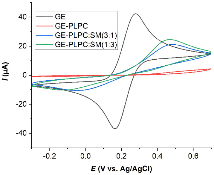
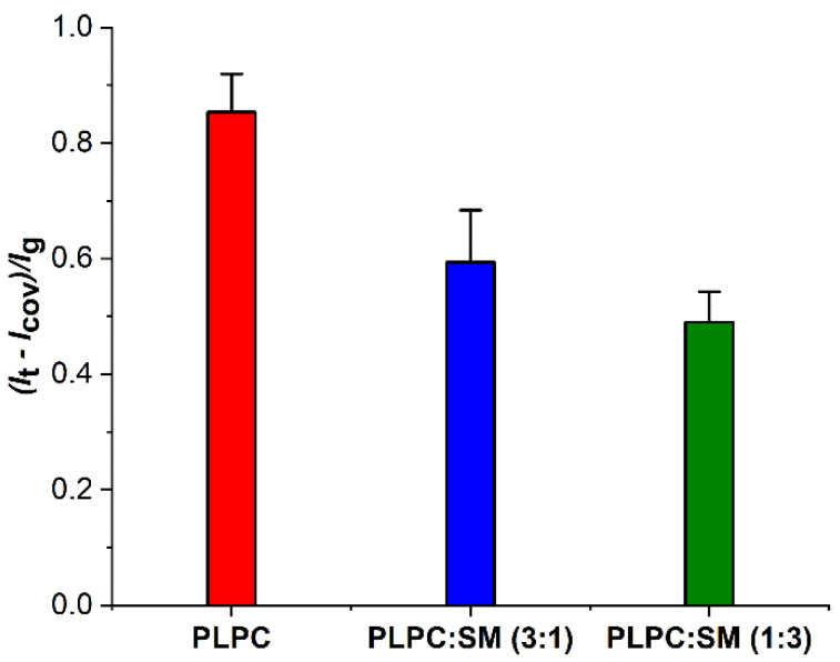
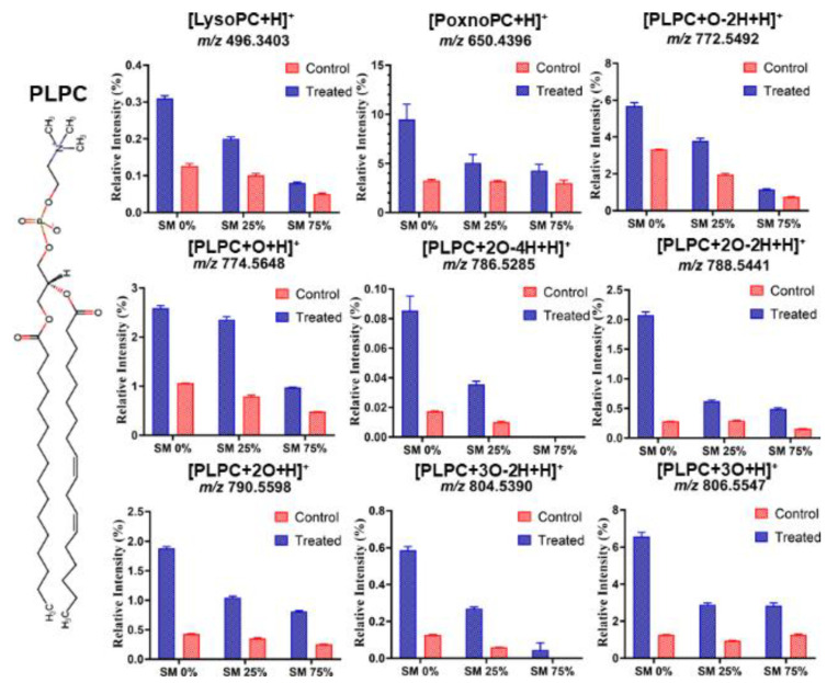
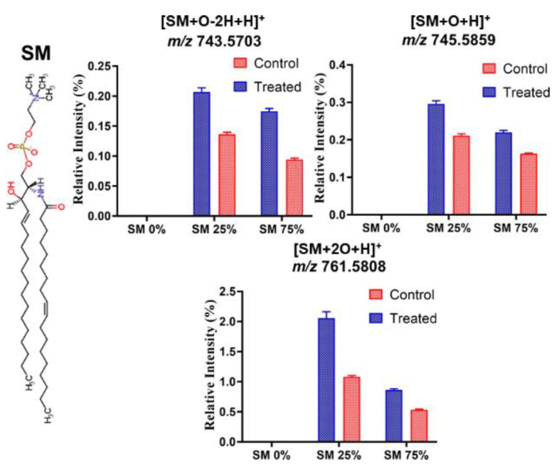
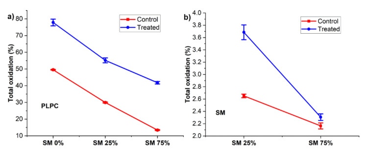
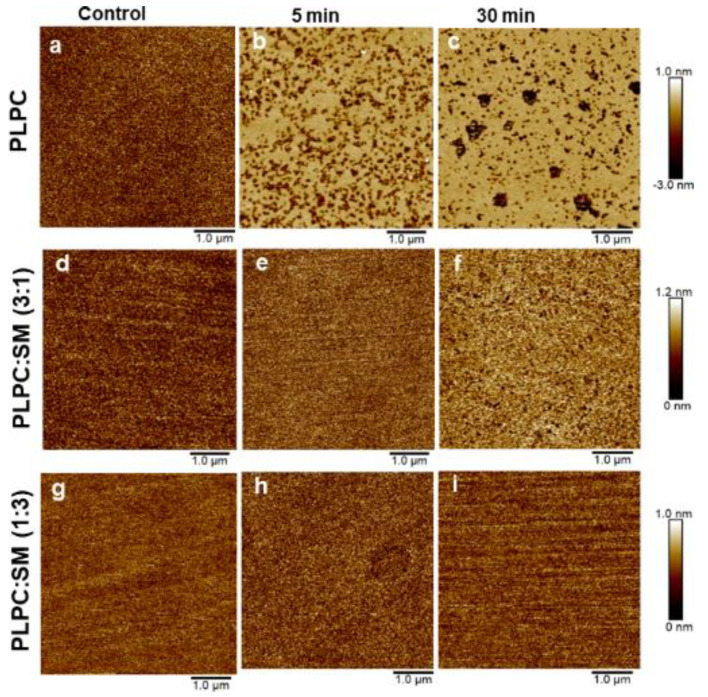
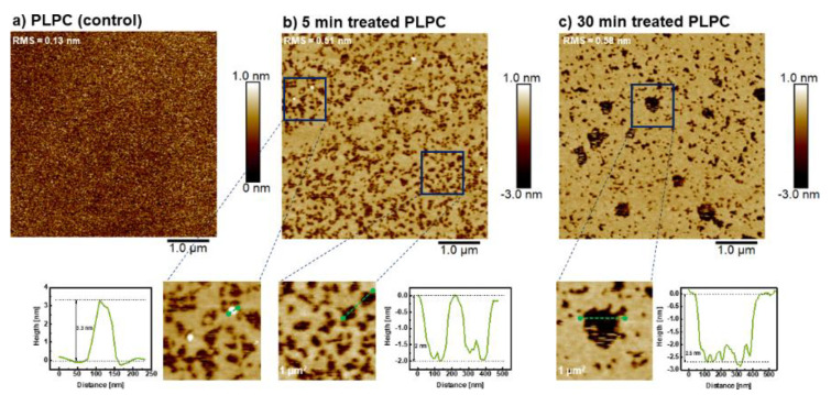
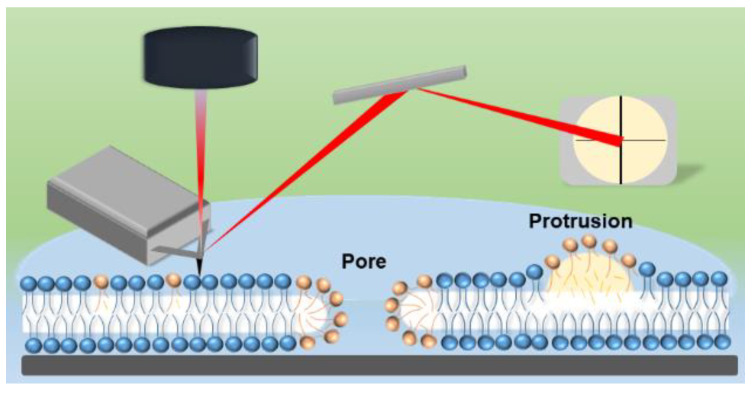
In the eye lens cell membrane, the lipid composition changes during the aging process: the proportion of sphingomyelins (SM) increases, that of phosphatidylcholines decreases. To investigate the protective role of the SMs in the lens cell membrane against oxidative damage, analytical techniques such as electrochemistry, high-resolution mass spectrometry (HR-MS), and atomic force microscopy (AFM) were applied. Supported lipid bilayers (SLB) were prepared to mimic the lens cell membrane with different fractions of PLPC/SM (PLPC: 1-palmitoyl-2-linoleoyl-sn-glycero-3-phosphocholine). The SLBs were treated with cold physical plasma. A protective effect of 30% and 44% in the presence of 25%, and 75% SM in the bilayer was observed, respectively. PLPC and SM oxidation products were determined via HR-MS for SLBs after plasma treatment. The yield of fragments gradually decreased as the SM ratio increased. Topographic images obtained by AFM of PLPC-bilayers showed SLB degradation and pore formation after plasma treatment, no degradation was observed in PLPC/SM bilayers. The results of all techniques confirm the protective role of SM in the membrane against oxidative damage and support the idea that the SM content in lens cell membrane is increased during aging in the absence of effective antioxidant systems to protect the eye from oxidative damage and to prolong lens transparency.

Keywords: aging; atomic force microscopy; cold physical plasma; electrochemistry; eye lens cell membrane; mass spectrometry; oxidized lipids; sphingomyelin.


Conflict of interest statement

The authors declare no competing interests.

Figures

Figure 1
Figure 1
Cyclic voltammograms (CVs) of 10 mM K4[Fe(CN)6] in 50 mM phosphate buffer solution (scan rate 50 mV·s−1) for bare gold electrode (GE, black curve) and after formation of PLPC (PLPC, red curve), PLPC:SM (3:1, blue curve), and PLPC:SM (1:3, green curve) lipid bilayers, respectively.
Figure 2
Figure 2
Normalized recovery peak currents of ferrocyanide/ferricyanide redox system determined by differential pulse voltammetry of lipid bilayers on gold electrodes before (Icov) and after 30 min plasma treatment (It) (n = 3). Varied is the SM fraction in PLPC bilayers. Each system is normalized with respect to the peak current of the redox system for the bare gold electrode Ig.
Figure 3
Figure 3
The relative intensity for different PLPC oxidation products obtained from lipid bilayers with different SM fraction after 30 min plasma treatment (Treated) and after auto-oxidation (Control) (n = 3). Data were obtained by HR-mass spectroscopy. The relative intensity of each lipid peroxidation product (LPP) is calculated as the peak area of LPP divided by the peak area of unmodified PLPC in the same sample. Note that the different graphs have different scaling of the y-axis. 1-Palmitoyl-2-hydroxy-sn-glycero-3-phosphocholine and 1-palmitoyl-2-(9′-oxo-nonanoyl)-sn-glycero-3-phosphocholine were presented as LysoPC and PoxnoPC, respectively.
Figure 4
Figure 4
The relative intensity for different SM oxidation products obtained from lipid bilayers with different SM fraction after 30 min plasma treatments (Treated) and after auto oxidation (Control) (n = 3). Relative intensity of each lipid peroxidation product (LPP) is calculated as the peak area of LPP divided by the peak area of unmodified SM in the same sample. Note that the different graphs have different scaling of the y-axis.
Figure 5
Figure 5
Total oxidation of PLPC (a) and SM (b) after 30 min plasma treatment of lipid bilayers with different SM fraction (indicated “Treated”) and without treatment (indicated “Control”) (n = 3). Total oxidation is defined as the sum of lipid peroxidation product (LPP) peak areas quantified in each sample relative to the peak area of non-oxidized parent lipid. Note that the different graphs have different scaling of the y-axis.
Figure 6
Figure 6
AFM images (5.0 × 5.0 µm2) of supported PLPC bilayers with different SM fractions before (left) and after plasma treatment for different duration (centre 5 min; right 30 min). Measurements were performed in 50 mM phosphate buffer solution at room temperature. Note the different height scale; the assignment of the height scales is (a, d, e, g, h, i: 0 to 1 nm), (b, c: −3 to 1 nm) and (f: 0 to 1.2 nm).
Figure 7
Figure 7
AFM images of supported PLPC bilayers before (a), after 5 min; and (b) after 30 min; (c) plasma treatment. Bottom row: The images are magnifications of the indicated areas. Histograms along the green lines show the height of the protrusions (left), and the increasing depth of the pores with increasing treatment time. Experimental conditions as in Figure 6.
Figure 8
Figure 8
Schematic of the experimental AFM setup and supported PLPC bilayers after plasma treatment. Shown are both a pore and a protrusion. Brown color indicates fragmented or oxidized PLPC molecules, blue color the native state.

References

    1. Babizhayev M.A., Yegorov Y.E. Reactive oxygen species and the aging eye: Specific role of metabolically active mitochondria in maintaining lens function and in the initiation of the oxidation-induced maturity onset cataract—A novel platform of mitochondria-targeted antioxidants with broad therapeutic potential for redox regulation and detoxification of oxidants in eye diseases. Am. J. Ther. 2016;23:e98–e117. doi: 10.1097/MJT.0b013e3181ea31ff. - DOI - PubMed
    1. Flatt T. A new definition of aging? Front. Genet. 2012;3:148. doi: 10.3389/fgene.2012.00148. - DOI - PMC - PubMed
    1. Liguori I., Russo G., Curcio F., Bulli G., Aran L., Della-Morte D., Gargiulo G., Testa G., Cacciatore F., Bonaduce D., et al. Oxidative stress, aging, and diseases. Clin. Interv. Aging. 2018;13:757–772. doi: 10.2147/CIA.S158513. - DOI - PMC - PubMed
    1. Beckman K.B., Ames B.N. The free radical theory of aging matures. Physiol. Rev. 1998;78:547–581. doi: 10.1152/physrev.1998.78.2.547. - DOI - PubMed
    1. Truscott R.J. Age-related nuclear cataract-oxidation is the key. Exp. Eye Res. 2005;80:709–725. doi: 10.1016/j.exer.2004.12.007. - DOI - PubMed

Publication types

LinkOut - more resources