Review
. 2021 Feb 13;13(2):141.
doi: 10.3390/toxins13020141.

Distribution, Ecology, Chemistry and Toxicology of Plant Stinging Hairs

Affiliations
Review

Distribution, Ecology, Chemistry and Toxicology of Plant Stinging Hairs

Hans-Jürgen Ensikat et al. Toxins (Basel). .

Abstract

Plant stinging hairs have fascinated humans for time immemorial. True stinging hairs are highly specialized plant structures that are able to inject a physiologically active liquid into the skin and can be differentiated from irritant hairs (causing mechanical damage only). Stinging hairs can be classified into two basic types: Urtica-type stinging hairs with the classical "hypodermic syringe" mechanism expelling only liquid, and Tragia-type stinging hairs expelling a liquid together with a sharp crystal. In total, there are some 650 plant species with stinging hairs across five remotely related plant families (i.e., belonging to different plant orders). The family Urticaceae (order Rosales) includes a total of ca. 150 stinging representatives, amongst them the well-known stinging nettles (genus Urtica). There are also some 200 stinging species in Loasaceae (order Cornales), ca. 250 stinging species in Euphorbiaceae (order Malphigiales), a handful of species in Namaceae (order Boraginales), and one in Caricaceae (order Brassicales). Stinging hairs are commonly found on most aerial parts of the plants, especially the stem and leaves, but sometimes also on flowers and fruits. The ecological role of stinging hairs in plants seems to be essentially defense against mammalian herbivores, while they appear to be essentially inefficient against invertebrate pests. Stinging plants are therefore frequent pasture weeds across different taxa and geographical zones. Stinging hairs are usually combined with additional chemical and/or mechanical defenses in plants and are not a standalone mechanism. The physiological effects of stinging hairs on humans vary widely between stinging plants and range from a slight itch, skin rash (urticaria), and oedema to sharp pain and even serious neurological disorders such as neuropathy. Numerous studies have attempted to elucidate the chemical basis of the physiological effects. Since the middle of the 20th century, neurotransmitters (acetylcholine, histamine, serotonin) have been repeatedly detected in stinging hairs of Urticaceae, but recent analyses of Loasaceae stinging hair fluids revealed high variability in their composition and content of neurotransmitters. These substances can explain some of the physiological effects of stinging hairs, but fail to completely explain neuropathic effects, pointing to some yet unidentified neurotoxin. Inorganic ions (e.g., potassium) are detected in stinging hairs and could have synergistic effects. Very recently, ultrastable miniproteins dubbed "gympietides" have been reported from two species of Dendrocnide, arguably the most violently stinging plant. Gympietides are shown to be highly neurotoxic, providing a convincing explanation for Dendrocnide toxicity. For the roughly 648 remaining stinging plant species, similarly convincing data on toxicity are still lacking.

Keywords: Dendrocnide; Loasaceae; Tragia; Urtica; acetylcholine; defense mechanisms; herbivores; histamine; neurotransmitters; toxicity.


Conflict of interest statement

The authors declare no conflict of interest.

Figures
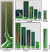
Figure 1
Figure 1
Size comparison of different Urtica type stinging hairs: Compositional-contrast scanning electron microscope (SEM) images of fresh or frozen hydrated (cryo-SEM) stinging hairs from different plant families. Euphorbiaceae: (a) Cnidoscolus aconitifolius. Urticaceae: (b) Urera baccifera, (c) Urtica mairei, (d) Urtica ferox, (e) Urtica atrovirens, and (f) Urtica dioica (arrow; one of the smallest ones). Loasaceae: (g) Nasa amaluzensis, (h) Caiophora deserticola, (i) Loasa insons, (j) Aosa rupestris, and (k) Chichicaste grandis. Caricaceae: (l) Horovitzia cnidoscoloides. Namaceae: (m) Wigandia ecuadorensis. Red color indicates mineralization with high concentrations of of Si or Ca. Scale bar = 1 mm.
Figure 2
Figure 2
Morphological details of stinging hairs of the Urtica type (including Wigandia). (ai) Urtica stinging hairs. Transmission light microscopy (LM) image of (a) entire hair and (b) section of basal part (SEM block face image) reveal the extent of the single stinging cell. SEM images of (c) critical-point- (CP-) dried and (d,e) air-dried stinging hairs demonstrate the rigidity of the strongly mineralized shaft in contrast to the flexibility of the shrinking non-mineralized tissue of the pedestal. (f) The function of a pipette imitates the ejection of fluid from a stinging hair. (g) LM image of the tip; the fluid-filled lumen extends into the bulb. (h,i) SEM images of intact and broken tip. (j,k) LM and SEM images of Wigandia stinging hair tips with slightly different shape, but also hollow up to the bulb. Scale bars: (a,c,e) 200 µm; (b) 100 µm; (d) 500 µm; (g,h,i) 20 µm; (j,k) 50 µm.
Figure 3
Figure 3
Morphological and chemical details of Tragia-type stinging hairs on Dalechampia aristolochiifolia plants. (a) Cryo-SEM image. (b,c) LM images, showing an optically dense (or intransparent) structure in the center of the apical part. (d) Raman spectrum of the dense structure in the tip; the peaks match perfectly with those of calciumoxalate-monohydrate. (e) Topographic image and calcium distribution image by EDX element mapping, showing high Ca concentration in apex. (f) EDX spectra of the locations marked in (e). (Image (a) Yaron Malkowsky, Nees Institut). Scale bars: (a,b) 50 µm; (c) 10 µm; (e) 100 µm.
Figure 4
Figure 4
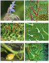
Irritant hairs. (a,b) Borago officinalis (Boraginaceae) showing (a) plant habit (flower) and (b) SEM image of leaf underside covered with pointed mineralized hairs. (c) Forskaolea angustifolia (Urticaceae) with mineralized, hook-shaped irritant trichomes. (d) Solanum carolinense (Solanaceae) with small, unmineralized, branched irritant trichomes; (e,f) Cucurbita pepo (Cucurbitaceae) showing (e) plant habit and (f) SEM image of multicellular mineralized hairs on leaf underside. (b,c,d,f) Compositional contrast cryo-SEM image. Own images except (d) Yaron Malkowsky, Nees Institute). Scale bars: (b) 500 µm; (c) 100 µm; (d,f) 200 µm.
Figure 5
Figure 5
Plant groups with stinging hairs. (a,b) Urtica dioica subsp. dioica (Urticaceae) female shoot of the particularly urticant “var. hispida” from the Swiss alps. (c) Caiophora deserticola (Loasaceae) flowering shoot. (d) Aosa rupestris (Loasaceae) young inflorescence. (e) Cnidoscolus liesneri (Euphorbiaceae). (f) Dalechamica aristolochiifolia (Euphorbiaceae) inflorescence. (g,h) Wigandia caracasana (Namaceae) showing (g) flowering shoot and (h) stem with dense cover of stinging hairs. (i) Horovitzia cnidoscoloides (Caricaceae) shoot apex. Own images, apart from (a) Bernadette Grosse-Veldmann, Nees Institut; (c) Markus Ackermann, Koblenz University; (f) Günther Gerlach, Botanischer Garten München-Nymphenburg; (g) Rafael Acuña, San José, CR; (h) Hartmut Hilger, Berlin.
Figure 6
Figure 6
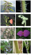
Stinging hair distribution on plants. (a) Urtica ferox (Urticaceae), young plant. (b) Urtica ferox (Urticaceae) stem and petioles. (c) Aosa rupestris (Loasaceae) principal vein abaxial. (d) Caiophora andina (Loasaceae) flower with dense stinging hair cover on petals. (e) C. deserticola stinging hairs on receptacle. (f) C. chuquitensis stinging hairs in ovary receptacle. (g) Nasa macrothyrsa (Loasaceae) with dense cover of stinging hairs (dark brown) on ovary. (h) Cnidoscolus quercifolius (Euphorbiaceae) young fruit with extremely dense cover of stinging hairs. (i) Tragia sp. (Euphorbiaceae) young fruit with extremely dense cover of stinging hairs. (j) Urtica dioica (Urticaceae) tepals on fruit with stinging hairs. Own images apart from (a,b) Nicolai M. Nürk, Heidelberg; (dg) Markus Ackermann, Koblenz; (h,i) Günther Gerlach, München. (j) False-color SEM image; scale bar = 500 µm.
Figure 7
Figure 7
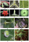
Homology of stinging hairs with other plant hairs. (a) Urtica dioica (Urticaceae) adaxial leaf surface with stinging hair (red, long pedestal, front left), shorter mineralized, unicellular trichomes (red, small pedestal), and minute glandular trichomes (spheroidal, green). (b) Caiophora lateritia (Loasaceae) with shorter pointed scabrid hairs and very small glochidiate hairs. (c) Loasa pallida (Loasaceae) with glochidiate and scabrid hairs on abaxial leaf surface. (d) Wigandia caracasana (Namaceae) with stinging hairs and shorter, stiff, unicellular trichomes on abaxial leaf surface. (e) Nama rothrockii (Namaceae) with stiff unicellular trichomes. (f) Phacelia malvifolia (Namaceae) with stiff, mineralized trichomes (red) and shorter, non-mineralized, glandular trichomes. Cryo-SEM compositional-contrast images, with red portion mineralized. Scale bars: (a,b) 500 µm; (cf) 200 µm.
Figure 8
Figure 8
Trichomes of particularly toxic representatives of Urticaceae. (a) Dendrocnide moroides stinging hairs, shorter stiff trichomes (both mineralized), and minute, spherical glandular trichomes. (bd) Urtica ferox, with (b) very long pedestal crowned with minute stinging hair, (c) stinging hair close-up, and (d) bulbous tip of stinging hair with pre-formed breakage point. Compositional-contrast SEM images, red portion mineralized. Scale bars: (a,c) 200 µm; (b) 500 µm; (d) 50 µm.
Figure 9
Figure 9
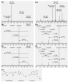
Nano-electrospray ionization mass spectra of stinging hair liquid of Loasa heterophylla (ac) and Caiophora deserticola (df). Assignments: 1 = histamine, 2 = serotonin, 3 = a monosaccharide C6H12O6. (a,d) Overview spectra, with only the low-mass region shown. Substances are identified via accurate mass and higher energy collision-induced dissociation (HCD) of mass-selected ions. (b) Fragmentation of m/z 112 from (a) spectrum in comparison to (c) fragmentation of a reference sample of histamine. (e) Fragmentation of m/z 177 from (d) spectrum in comparison to (f) fragmentation of a reference sample of serotonin. (g) Structure formulas of the neurotransmitters acetylcholine, histamine, and serotonin.
Figure 10
Figure 10
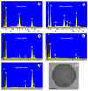
(ae) EDX element spectra of stinging hair fluid dry mass from various species. (f) SEM image of a dried droplet of Urera baccifera stinging hair fluid with dendritic KCl-crystal complexes. Scale bar = 200 µm.
Figure 11
Figure 11
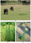
Stinging plants and herbivory. (a) Overgrazed pasture with ponies, with only Urtica dioica (Urticaceae) left standing (near Bonn, Germany). (b) Urtica urens (Urticaceae) in a sheep pasture; note closely cropped grass next to untouched nettle (Mooi River, RSA). (c) Urtica urens as only surviving plant in an enclosure for prairie dogs (Cynomys, Tierpark Friedrichsfelde, Berlin. Germany). Own images apart from (b) Alexandra Krühn, Berlin.
Figure 12
Figure 12
Stinging plants and herbivory. (a) Caterpillar feeding on leaves of Cnidoscolus (Euphorbiaceae, Northern Peru, Cajamarca); note latex seeping from leaf vein. (b) Poisonous latex in Cnidoscolus leaf stalk. (c) Caterpillar feeding on leaves of Wigandia sp. (Namaceae) in Peru. (d) Skeletized leaves of Nasa rubrastra (Loasaceae) in cloud forest (Ecuador, Tungurahua). (e) Nasa amaluzensis with brown stinging hairs and glandular hairs producing oily secretions. Own images apart from (d) Rafael Acuña, San Jose, Costa Rica.

References

    1. Thurston E.L., Lersten N.R. The morphology and toxicology of plant stinging hairs. Bot. Rev. 1969;35:393–412. doi: 10.1007/BF02858878. - DOI
    1. Hooke R. John Martyn “Printers to the Royal Society”. Dover Pub. Co.; New York, NY, USA: 1965. Micrographia: Or Some Physiological Descriptions of Minute Bodies Made by Magnifying Glasses with Observations and Inquiries thereupon.
    1. Mustafa A., Ensikat H.J., Weigend M. Stinging hair morphology and wall biomineralization across five plant families: Conserved morphology versus divergent cell wall composition. Am. J. Bot. 2018;105:1109–1122. doi: 10.1002/ajb2.1136. - DOI - PubMed
    1. Gorup-Besanetz E.F. Notiz über das Vorkommen der Ameisensäure in den Brennnesseln. J. Prakt. Chem. 1849;48:191–192. doi: 10.1002/prac.18490480124. - DOI
    1. Rauter J. Zur Entwicklungsgeschichte einiger Trichomgebilde. Denkschr. Akad. Wiss. 1872;31:2–49.

MeSH terms

LinkOut - more resources