Review
. 2021 Feb 13;13(2):142.
doi: 10.3390/toxins13020142.

Uremic Toxins in the Progression of Chronic Kidney Disease and Cardiovascular Disease: Mechanisms and Therapeutic Targets

Affiliations
Review

Uremic Toxins in the Progression of Chronic Kidney Disease and Cardiovascular Disease: Mechanisms and Therapeutic Targets

Yong Jin Lim et al. Toxins (Basel). .

Abstract

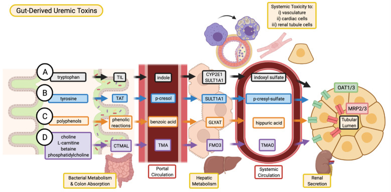
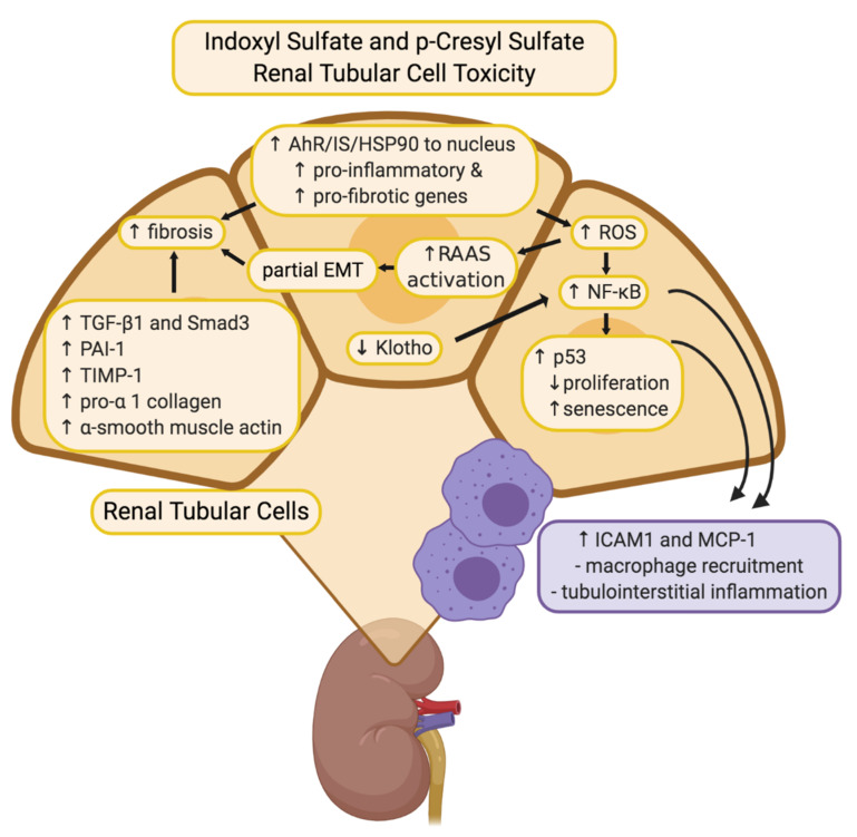
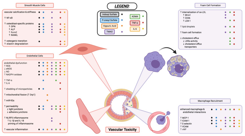
Chronic kidney disease (CKD) is a progressive loss of renal function. The gradual decline in kidney function leads to an accumulation of toxins normally cleared by the kidneys, resulting in uremia. Uremic toxins are classified into three categories: free water-soluble low-molecular-weight solutes, protein-bound solutes, and middle molecules. CKD patients have increased risk of developing cardiovascular disease (CVD), due to an assortment of CKD-specific risk factors. The accumulation of uremic toxins in the circulation and in tissues is associated with the progression of CKD and its co-morbidities, including CVD. Although numerous uremic toxins have been identified to date and many of them are believed to play a role in the progression of CKD and CVD, very few toxins have been extensively studied. The pathophysiological mechanisms of uremic toxins must be investigated further for a better understanding of their roles in disease progression and to develop therapeutic interventions against uremic toxicity. This review discusses the renal and cardiovascular toxicity of uremic toxins indoxyl sulfate, p-cresyl sulfate, hippuric acid, TMAO, ADMA, TNF-α, and IL-6. A focus is also placed on potential therapeutic targets against uremic toxicity.

Keywords: asymmetric dimethylarginine; cardiovascular disease; chronic kidney disease; hippuric acid; indoxyl sulfate; interleukin 6; p-cresyl sulfate; trimethylamine N-oxide; tumor necrosis factor al-pha; uremic toxins.


Conflict of interest statement

The authors declare no conflict of interest.

Figures

Figure 1
Figure 1
Absorption, and excretion pathway of gut-derived uremic toxins. (A) Indoxyl sulfate is produced when dietary tryptophan is converted into indole by bacterial tryptophan indole-lyase (TIL) and subsequent absorption into the portal circulation for further metabolism by cytochrome P450 2E1 (CYP2E1) and sulfotransferase 1A1 (SULT1A1). (B) P-cresyl sulfate begins as dietary tyrosine which is metabolized by tyrosine aminotransferase (TAT) into p-cresol. These intermediates are converted into p-cresyl sulfate through SULT1A1. (C) Various dietary polyphenols are converted through multiple phenolic reactions by colon bacteria into benzoic acid which is then conjugated with glycine by glycine-N-acyltransferase (GLYAT) to produce hippuric acid. (D) TMAO is the product of TMA oxidation by flavin-containing monooxygenase 3 (FMO3), where TMA is the intermediate metabolite of carnitine trimethylamine lyase (CTMAL) breakdown of various dietary molecules such as choline, L-carnitine, betaine, and phosphatidylcholine. (AD) Uremic toxins are secreted by renal tubular cell via drug transporters (organic anion transporter 1/3, OAT1/3; and multidrug resistance-associated proteins 2/3, MRP2/3 and subsequently excreted in the urine. Image created with BioRender.com.
Figure 2
Figure 2
Overview of putative mechanisms of renal toxicity for IS and pCS. Through various inflammatory and fibrotic pathways, both IS and pCS have been described to mediate toxicity to renal tubular cells in similar ways. Increased expression of various fibrotic genes such as TGF-β1, TIMP-1, pro-α1 (I) collagen contribute to alterations in the tubular cell morphology and structure of the ECM. Involvement of ROS and NF-κB reduce the proliferative capacity of tubular cells, contribute to partial EMT leading to fibrosis, and recruit macrophages to produce additional tubulointerstitial inflammation. Image created with BioRender.com.
Figure 3
Figure 3
Vascular toxicity of uremic toxins through impacts on endothelial cell function, vascular smooth muscle cell morphology, and macrophage recruitment and transformation into foam cells of atherosclerotic plaques. Each coloured dot (see below and legend in figure) represents a uremic toxin having evidence in the literature to contribute to the various processes, or changes in cell activity and function described. Indoxyl sulfate (●; black), p-cresyl sulfate (; blue), hippuric acid (; orange), TMAO (; purple), ADMA (; green), TNF-α (; red), and IL-6 (; yellow) are shown to have varying effects in the cell types and cellular processes described above. Endothelial dysfunction and inflammation, vascular calcification and stiffness, enhanced interactions between macrophages and endothelial cells, as well as formation of foam cells are critical components to the various uremic toxins’ vascular toxicities. Some toxins may contribute to these mechanisms of toxicity but have yet to be elucidated or confirmed in the literature; some toxins have contradicting evidence that needs further investigation. This figure does not necessarily describe the complex interplay between the uremic toxins, inflammatory markers, and immune system that likely play a large interconnected role in the vascular toxicity contributing to CVD. Image created with BioRender.com.

References

    1. Stevens P.E., Levin A. Evaluation and management of chronic kidney disease: Synopsis of the kidney disease: Improving global outcomes 2012 clinical practice guideline. Ann. Intern. Med. 2013;158:825–830. doi: 10.7326/0003-4819-158-11-201306040-00007. - DOI - PubMed
    1. Chen T.K., Knicely D.H., Grams M.E. Chronic Kidney Disease Diagnosis and Management: A Review. JAMA J. Am. Med. Assoc. 2019;322:1294–1304. doi: 10.1001/jama.2019.14745. - DOI - PMC - PubMed
    1. KDIGO Clinical Practice Guideline for the Evaluation and Management of Chronic Kidney Disease. Kidney Int. Suppl. 2013;3:1–150.
    1. Neovius M., Jacobson S.H., Eriksson J.K., Elinder C.G., Hylander B. Mortality in chronic kidney disease and renal replacement therapy: A population-based cohort study. BMJ Open. 2014;4:1–9. doi: 10.1136/bmjopen-2013-004251. - DOI - PMC - PubMed
    1. Gansevoort R.T., Correa-Rotter R., Hemmelgarn B.R., Jafar T.H., Heerspink H.J.L., Mann J.F., Matsushita K., Wen C.P. Chronic kidney disease and cardiovascular risk: Epidemiology, mechanisms, and prevention. Lancet. 2013;382:339–352. doi: 10.1016/S0140-6736(13)60595-4. - DOI - PubMed