. 2021 Feb 13;26(4):1000.
doi: 10.3390/molecules26041000.

Chemical Profile of Launaea nudicaulis Ethanolic Extract and Its Antidiabetic Effect in Streptozotocin-Induced Rats

Affiliations

Chemical Profile of Launaea nudicaulis Ethanolic Extract and Its Antidiabetic Effect in Streptozotocin-Induced Rats

Samah A El-Newary et al. Molecules. .

Abstract

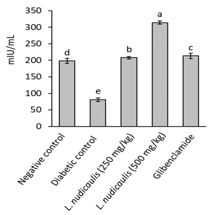
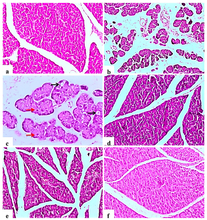
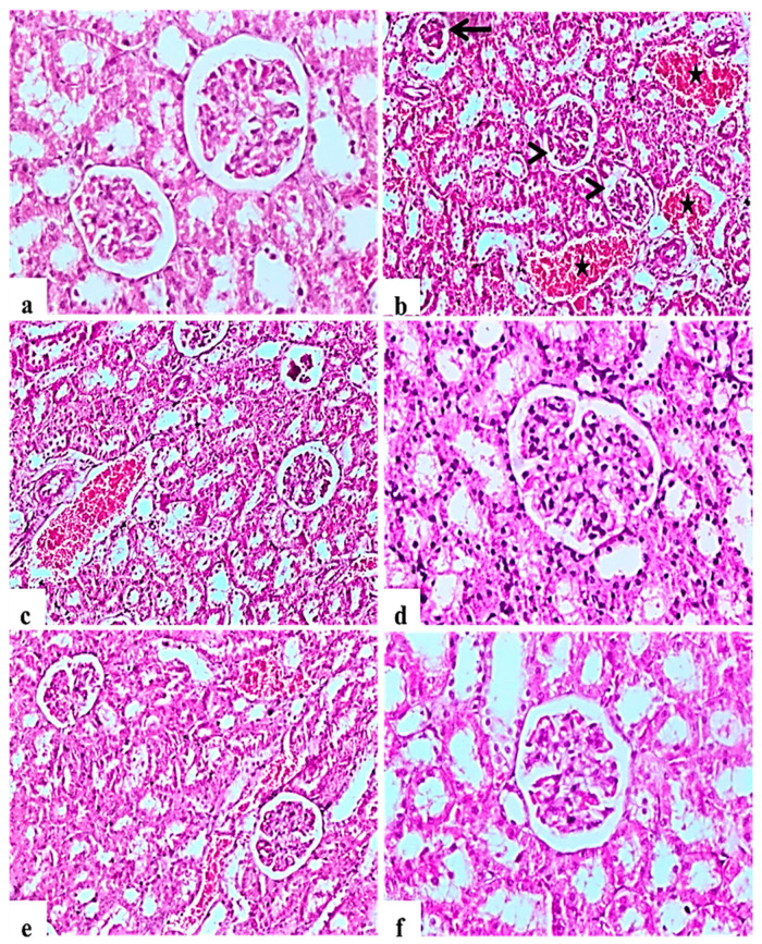
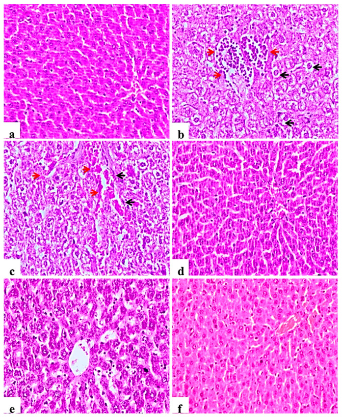
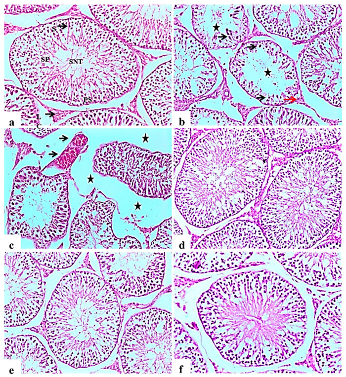
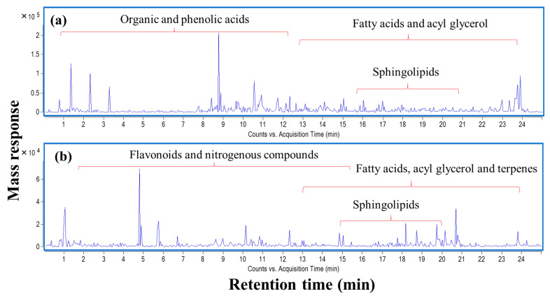
Launaea nudicaulis is used in folk medicine worldwide to treat several diseases. The present study aimed to assess the antidiabetic activity of L. nudicaulis ethanolic extract and its effect on diabetic complications in streptozotocin-induced hyperglycemic rats. The extract was orally administrated at 250 and 500 mg/kg/day for 5-weeks and compared to glibenclamide as a reference drug at a dose of 5 mg/kg/day. Administration of the extract exhibited a potential hypoglycemic effect manifested by a significant depletion of serum blood glucose concurrent with a significant elevation in serum insulin secretion. After 5-weeks, extract at 250 and 500 mg/kg/day decreased blood glucose levels by about 53.8 and 68.1%, respectively, compared to the initial values (p ≤ 0.05). The extract at the two dosages prevented weight loss of rats from the 2nd week till the end of the experiment, compared to diabetic control rats. The extract further exhibited marked improvement in diabetic complications including liver, kidney and testis performance, oxidative stress, and relative weight of vital organs, with respect to diabetic control. Histopathological examinations confirmed the previous biochemical analysis, where the extract showed a protective effect on the pancreas, liver, kidney, and testis that degenerated in diabetic control rats. To characterize extract composition, UPLC-ESI-qTOF-MS identified 85 chromatographic peaks belonging to flavonoids, phenolics, acyl glycerols, nitrogenous compounds, and fatty acids, with four novel phenolics reported. The potential anti-diabetic effect warrants its inclusion in further studies and or isolation of the main bioactive agent(s).

Keywords: LCMS; Launaea nudicaulis; antihyperglycemic; histopathological studies; liver and kidney functions; metabolites profiling.


Conflict of interest statement

The authors declare no conflict of interest.

Figures

Figure 1
Figure 1
Insulin concentration of STZ- induced diabetic rats treated with L. nudicaulis ethanolic extract during 5-weeks. Data are presented as the means ± SE (n = 10). Data analyzed by ANOVA one-way. Value with the different superscript letters means significance at a probability level of 0.05.
Figure 2
Figure 2
Light photomicrographs of pancreatic tissue (H&E ×100). (a): control rat showing normal architecture of the pancreas, acinar cells and arranged into small lobules, pancreatic lobules separated by intact intralobular and interlobular connective tissue septa. (b): Subgroup I (diabetic control) showing disorganization of the structure of the endocrine and exocrine cells, shrunken islets with a drastic decrease in the number, necrosis in Langerhans islets, acinar cells were swollen and vacuolated. (c): Some islet cells have degenerated and vacuolated (Black arrows), cellular apoptosis (Red arrows), in the form of small pyknotic nuclei and deeply acidophilic cytoplasm (×200). (d): Subgroup IV (STZ treated diabetic-rat + glibenclamide) showing normal pancreatic structures and exocrine acini surrounding the islets of Langerhans. (e): Subgroup II (STZ treated diabetic-rat + 250 mg ext./kg/day) showing moderate necrotic changes of the pancreas associated with mild reduction in the size and number of the islets of Langerhans. (f): Subgroup III (STZ treated diabetic-rat + 500 mg ext./kg/day) showing a remarkable recovery as compared to the STZ treated rats, with mild degree of necrosis of islets of Langerhans.
Figure 3
Figure 3
Histological H&E stained sections of Kidney (ae) (×200) (b,d) (×100). a: control untreated rat showing normal glomerulus and renal tubules. b and c: Subgroup I (STZ treated diabetic-rat) rats (STZ treated diabetic-rat) showing atrophy of glomerular tuft (Black arrow), degeneration of renal tubules and increase in the Bowman’ space area (Arrow heads) associated with severe hemorrhage and congestion of renal blood vessels (stars). (d): Subgroup IV (STZ treated diabetic-rat + glibenclamide) rats marked amelioration of the glomerular and tubular damage. (e): Subgroup II (STZ treated diabetic-rat + 250 mg ext./kg/day) rats showing mild recovery of the pathological picture, still with mild congestion of renal blood vessels. (f): Subgroup III (STZ treated diabetic-rat + 500 mg ext./kg/day) rats showing marked recovery and amelioration of the pathological picture associated with the restoration of normal renal architecture.
Figure 4
Figure 4
Histological H&E stained sections of liver (×200). (a): normal central veins; hepatocytes with the blood sinusoids are present between the hepatic cords. (b): Subgroup I (STZ treated diabetic-rat) rats showing mononuclear cell infiltration of the hepatic tissue (Red arrow), Kupffer cell activation, severe vacuolar hydropic degeneration of hepatocytes (Black arrow). (c): G1 rats showing Kupffer cell activation (Black arrow), severe vacuolar hydropic degeneration of hepatocytes with severe congestion of central veins and blood sinusoids (red arrow). (d): Subgroup IV (STZ treated diabetic-rat + glibenclamide) showing almost normal morphology. (e): Subgroup II (STZ treated diabetic-rat + 250 mg ext./kg/day) showing less improvement or restoration of hepatocyte degeneration. (f): Subgroup III (STZ treated diabetic-rat + 500 mg ext./kg/day) showing normal architecture with mild vascular congestion.
Figure 5
Figure 5
Histological H&E stained sections of the testis (×100). (a): control rat showing normal seminiferous tubules architecture (SNT), showing a clear lumen and a normal arrangement of cellular types, lined with series of spermatogenic cells; spermatogonia, primary spermatocytes (PS) and spermatids. Sertoli cells (S) are seen with attached sperms (SP). Interstitial space containing normal Leydig cells (LC). (b): Subgroup I (STZ treated diabetic-rat rats showing disorganization and atrophy of seminiferous tubules, absence of spermatogenesis (Star) and severe degenerative change of the lining epithelium Sertoli cells (black arrow). Leydig cells atrophy and vacuolations (red arrow). (c): showing disorganization and atrophy of seminiferous tubules, absence of spermatogenesis associated with severe interstitial oedema (Stars) and severe congestion of testicular blood vessels (black arrow). (d), (e,f): Subgroup IV (STZ treated diabetic-rat + glibenclamide, Subgroup II (STZ treated diabetic-rat + 250 mg ext./kg/day) and Subgroup III (STZ treated diabetic-rat + 500 mg ext./kg/day), respectively showing normal structure of seminiferous tubules with mild interstitial oedema, associated with the restoration of spermatogenic cells in most seminiferous tubules.
Figure 6
Figure 6
UPLC-qTOF-MS base peak chromatogram of L. nudicaulis ethanolic extract detected in (a) negative and (b) positive ionization modes.

References

    1. Saeedi P., Petersohn I., Salpea P., Malanda B., Karuranga S., Unwin N., Colagiuri S., Guariguata L., Motala A.A., Ogurtsova K. Global and regional diabetes prevalence estimates for 2019 and projections for 2030 and 2045: Results from the International Diabetes Federation Diabetes Atlas. Diabetes Res. Clin. Pract. 2019;157:107843. doi: 10.1016/j.diabres.2019.107843. - DOI - PubMed
    1. Thomas R., Halim S., Gurudas S., Sivaprasad S., Owens D. IDF Diabetes Atlas: A review of studies utilising retinal photography on the global prevalence of diabetes related retinopathy between 2015 and 2018. Diabetes Res. Clin. Pract. 2019;157:107840. doi: 10.1016/j.diabres.2019.107840. - DOI - PubMed
    1. Osawa T., Kato Y. Protective role of antioxidative food factors in oxidative stress caused by hyperglycemia. Ann. N. Y. Acad. Sci. 2005;1043:440–451. doi: 10.1196/annals.1333.050. - DOI - PubMed
    1. Yin P., Zhao S., Chen S., Liu J., Shi L., Wang X., Liu Y., Ma C. Hypoglycemic and hypolipidemic effects of polyphenols from burs of Castanea mollissima Blume. Molecules. 2011;16:9764–9774. doi: 10.3390/molecules16119764. - DOI - PMC - PubMed
    1. Abdel-Haleem S.A., Ibrahim A.Y., Ismail R.F., Shaffie N.M., Hendawy S., Omer E. In-vivo hypoglycemic and hypolipidemic properties of Tagetes lucida alcoholic extract in streptozotocin-induced hyperglycemic Wistar albino rats. Ann. Agric. Sci. 2017;62:169–181. doi: 10.1016/j.aoas.2017.11.005. - DOI

MeSH terms

LinkOut - more resources