Review
. 2021 Feb 25;3(1):189-226.
doi: 10.3390/clockssleep3010012.

Circadian Rhythms of the Hypothalamus: From Function to Physiology

Affiliations
Review

Circadian Rhythms of the Hypothalamus: From Function to Physiology

Rachel Van Drunen et al. Clocks Sleep. .

Abstract

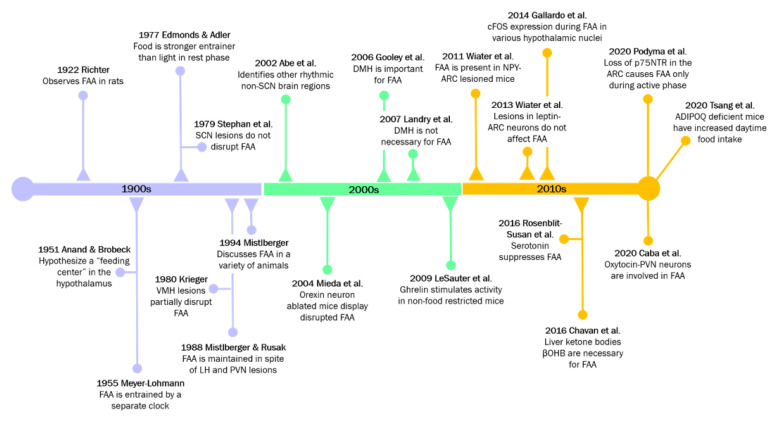
The nearly ubiquitous expression of endogenous 24 h oscillations known as circadian rhythms regulate the timing of physiological functions in the body. These intrinsic rhythms are sensitive to external cues, known as zeitgebers, which entrain the internal biological processes to the daily environmental changes in light, temperature, and food availability. Light directly entrains the master clock, the suprachiasmatic nucleus (SCN) which lies in the hypothalamus of the brain and is responsible for synchronizing internal rhythms. However, recent evidence underscores the importance of other hypothalamic nuclei in regulating several essential rhythmic biological functions. These extra-SCN hypothalamic nuclei also express circadian rhythms, suggesting distinct regions that oscillate either semi-autonomously or independent of SCN innervation. Concurrently, the extra-SCN hypothalamic nuclei are also sensitized to fluctuations in nutrient and hormonal signals. Thus, food intake acts as another powerful entrainer for the hypothalamic oscillators' mediation of energy homeostasis. Ablation studies and genetic mouse models with perturbed extra-SCN hypothalamic nuclei function reveal their critical downstream involvement in an array of functions including metabolism, thermogenesis, food consumption, thirst, mood and sleep. Large epidemiological studies of individuals whose internal circadian cycle is chronically disrupted reveal that disruption of our internal clock is associated with an increased risk of obesity and several neurological diseases and disorders. In this review, we discuss the profound role of the extra-SCN hypothalamic nuclei in rhythmically regulating and coordinating body wide functions.

Keywords: circadian rhythm; clock genes; extra-SCN hypothalamic nuclei; food-entrainable oscillator; hypothalamus; metabolism; obesity.


Conflict of interest statement

The authors declare no conflict of interest.

Figures

Figure 1
Figure 1
Interactions between the core clock and intracellular metabolism. The heterodimerization of BMAL1 and CLOCK proteins and subsequent binding to E-Box-containing regulatory elements leads to expression of the repressor PER and CRY proteins, the REV-ERBs and RORs, which initiate the auxiliary loop, and the core clock genes (CCGs) that drive numerous other intracellular rhythms. Cytoplasmic PER and CRY proteins are eventually tagged for degradation by AMPK. Rhythmic cellular metabolism, such as rhythmic NAD+ abundance, participates in the core clock by direct regulation of clock-associated factors, such as the anti-aging-associated histone deacetylase protein, SIRT1.
Figure 2
Figure 2
A circuit of hypothalamic oscillators. Numerous afferent and efferent projections characterize the hypothalamic landscape. The “master clock”, or the SCN, projects to the major hypothalamic nuclei while only a few nuclei project back to the SCN. Some regions, such as the DMH and ARC host an autonomous clock while other regions such as the PVN, LH and MPOA/VLPO are more heavily dependent on rhythmic innervations. Clock autonomy for the VMH and SPVZ has not been shown. An array of neural subpopulations in the hypothalamus are sensitive to hormones such as leptin, ghrelin, cholecystokinin (CCK), glucose and insulin, which cross the blood–brain barrier (BBB). These hypothalamic nuclei are often characterized by their inhibitory glutamatergic (blue), or their excitatory GABAergic (orange) projections. The gray dotted vertical lines denote the different regions of the hypothalamus. The general flow of information progresses from anterior to posterior, as indicated by the black arrow. Gastrin-Releasing Peptide; TRH = tyrosine hydroxylase; TRPM2 = transient receptor potential cation channel, subfamily M, member 2; VIP = vasoactive intestinal peptide.
Figure 3
Figure 3
Tracking the “food-entrainable oscillator” through time.
Figure 4
Figure 4
Organism-wide interactions with hypothalamic nuclei alter metabolism and energy homeostasis. AMPK activity is inversely affected by fasting, ghrelin and hypoglycemia compared to insulin, feeding, GLP-1 and leptin. Inhibition of mTORC1 by AMPK increases Agrp, in turn inhibiting MC4R neurons, and promoting energy intake. Alternately, inhibition of AMPK allows mTORC1 activity, which promotes pomc expression, oxidative metabolism, and an increase in activation of the MC4R-PVN neurons that signal satiety. In addition, the activation of MC4R neurons stimulates production of eCB, which feedback to POMC neurons to increase glutamate production, thereby increasing glutamatergic POMC neuronal input to the PVN. Nutrient stress is also a strong inhibitor of mTORC1 activity and a stimulator of SIRT1 expression in POMC-ARC neurons, whose downstream projections initiate WAT browning. Fasting results in AMPK activity in CRH-PVN neurons, resulting in a carbohydrate preference during re-feeding. Other nutrient signals such as insulin also stimulate the SF1-VMH neurons to express SIRT1 that increases insulin insensitivity in skeletal muscle. The thyroid hormone, T3 elicits AMPK activity in the SF1-VMH neurons and through a dual pathway that increases JNK1 and decreases ER stress to produce hepatic lipogenesis and BAT thermogenesis/WAT browning, respectively.
Figure 5
Figure 5
Hypothalamic-mediated circadian regulation of physiological functions. Powerful zeitgebers such as light and food have extensive effects on the circadian activity of cellular and tissue functions. The light-sensitive ipRGCs in the retina send signals to the master clock, the SCN, via the RHT. Food consumption stimulates various metabolic and digestive mechanisms which produce a variety of molecular signals and hormones. Nutrient-sensitive neurons in the circadian-regulated hypothalamus pick up the fluctuations in nutrients and hormones and relay the information downstream to the PB and the NTS which regulate the function of tissue processes through the parasympathetic and the sympathetic nervous system (PNS/SNS). With the exception of the intestines, which currently have not been found to have direct innervation from any hypothalamic nuclei, many metabolic-regulating tissues have direct or indirect innervations from various hypothalamic nuclei.

Similar articles

Cited by

References

    1. Takahashi J.S. Transcriptional architecture of the mammalian circadian clock. Nat. Rev. Genet. 2017;18:164–179. doi: 10.1038/nrg.2016.150. - DOI - PMC - PubMed
    1. Partch C.L., Green C.B., Takahashi J.S. Molecular architecture of the mammalian circadian clock. Trends Cell Biol. 2014;24:90–99. doi: 10.1016/j.tcb.2013.07.002. - DOI - PMC - PubMed
    1. Crumbley C., Wang Y., Kojetin D.J., Burris T.P. Characterization of the core mammalian clock component, NPAS2, as a REV-ERBα/RORα target gene. J. Biol. Chem. 2010;285:35386–35392. doi: 10.1074/jbc.M110.129288. - DOI - PMC - PubMed
    1. Gerhart-Hines Z., Feng D., Emmett M.J., Everett L.J., Loro E., Briggs E.R., Bugge A., Hou C., Ferrara C., Seale P., et al. The nuclear receptor Rev-erbα controls circadian thermogenic plasticity. Nature. 2013;503:410–413. doi: 10.1038/nature12642. - DOI - PMC - PubMed
    1. Kornmann B., Schaad O., Bujard H., Takahashi J.S., Schibler U. System-driven and oscillator-dependent circadian transcription in mice with a conditionally active liver clock. PLoS Biol. 2007;5:e34. doi: 10.1371/journal.pbio.0050034. - DOI - PMC - PubMed

LinkOut - more resources