Review
. 2021 Feb 25;13(3):358.
doi: 10.3390/v13030358.

Imaging Techniques to Study Plant Virus Replication and Vertical Transmission

Affiliations
Review

Imaging Techniques to Study Plant Virus Replication and Vertical Transmission

María Amelia Sánchez Pina et al. Viruses. .

Abstract

Plant viruses are obligate parasites that need to usurp plant cell metabolism in order to infect their hosts. Imaging techniques have been used for quite a long time to study plant virus-host interactions, making it possible to have major advances in the knowledge of plant virus infection cycles. The imaging techniques used to study plant-virus interactions have included light microscopy, confocal laser scanning microscopy, and scanning and transmission electron microscopies. Here, we review the use of these techniques in plant virology, illustrating recent advances in the area with examples from plant virus replication and virus plant-to-plant vertical transmission processes.

Keywords: correlative microscopy; electron microscopy; imaging; immunocytochemistry; in situ hybridization; light microscopy; plant virus–host interactions; viral life cycle; viral replication; viral transmission.


Conflict of interest statement

The authors declare no conflict of interest.

Figures

Figure 1
Figure 1
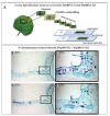
Analysis of the spatial distribution of a virus in infected plants by tissue printing hybridization. (A) Schematic representation of the tissue printing hybridization technique. Frequently after sectioning, the organ surface is printed onto a positively charged nylon or nitrocellulose membrane. Macromolecules, including viral nucleic acids, are transferred from the tissue to the membrane. The blotted membrane is then incubated with a digoxigenin (DIG)-labeled nucleic acid probe and the target nucleic acid is detected after incubation with a conjugated anti-DIG antibody and the appropriated chemiluminescent substrate. (BG) Tissue print hybridization using a digoxigenin-labelled RNA probe for detecting melon necrotic spot virus (MNSV) vRNA in (B) the first systemic leaves (developed leaves), (C) hypocotyl and main stem, (D) shoot tip, (E) second systemic leaf (young leaf), (F) inoculated cotyledon, and (G) roots of MNSV-infected melon plants. Insets displayed in B, E, and F corresponds to longitudinal or cross-sectional printings of the petioles. This figure is adapted with permission from [8].
Figure 2
Figure 2
In situ hybridization (ISH) assay on consecutive serial cross sections of Nicotiana benthamiana leaves infected with two isolates belonging to different strains of pepino mosaic virus (PepMV-EU and PepMV-CH2). (A) Schematic representation of the ISH assay. Small pieces of mock and mixed infected N. benthamiana leaves were fixed with paraformaldehyde (PFA), embedded into paraffin and sectioned. The ISH was performed on consecutive leaf cross sections of the same sample by using each probe (I or II) in each slide (1 or 2), respectively. (B,D) Images of the ISH performed on a pair of consecutive leaf serial cross sections from mixed (PepMV-EU + PepMV-CH2) inoculated plants with either the riboprobe for PepMV-EU (B) or PepMV-CH2; both viruses are located in dark-blue colored areas of the leaf sections distributed in patches of infected tissue, mixed with areas of noninfected tissue. (C,E) Higher magnification of the area boxed in B, D, respectively. The viral RNAs of both PepMV-EU and PepMV-CH2 isolates are located in the same dark-blue colored cells (arrowhead) of the leaf, mixed together with other noninfected tissue cells, or only infected by one of the isolates. Scale bars are displayed in the images. This figure is adapted with permission from [16].
Figure 3
Figure 3
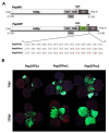
Pepino mosaic virus (PepMV) vectors expressing the green fluorescent protein (GFP). (A) Schematic representation (not to scale) of the PepMV genome and modified variants PepGFP2a, PepGFPm1, PepGFPm2, carrying the GFP gene. The GFP was expressed as a fusion to the coat protein (CP) through the foot and mouth disease virus 2A catalytic peptide sequence. The nucleotides marked in red correspond to synonymous mutations introduced into the corresponding vector versions to avoid sequence duplications. (B) Nicotiana benthamiana plants infected with PepMV vectors expressing GFP. Fluorescence was visualized in plants inoculated with PepGFP2a, PepGFPm1 or PepGFPm2 under UV light at 7 and 12 d post-inoculation (dpi). This figure is adapted with permission from [58].
Figure 4
Figure 4
Transmission electron microscopy study of melon necrotic spot virus (MNSV)-infected cells from melon cotyledons. (A) Altered mitochondria (M) where the replication of the virus takes place, showing the presence of small vesicles (black arrow) inside large dilations (star) and around the periphery of the organelle. (B) Detail of a big dilation surrounded by bottlenecked vesicles connecting either with the dilation lumen or with the cytoplasm. (C) MNSV-altered mitochondria in the proximity of plasmodesmata. (D) Ultrastructure of a healthy plant mitochondria. Samples were taken 3 days post-inoculation. Notes: Chl, chloroplast; CW, cell wall; Cyt, cytoplasm; M, mitochondria; Pd, plasmodesmata. Scale bars are displayed with the images.
Figure 5
Figure 5
Transmission electron microscopy (TEM) analysis and 3D reconstruction of melon necrotic spot virus (MNSV)-altered mitochondria by focused ion beam/field emission scanning electron microscopy (FIB-FESEM). (A) TEM image of the samples used for FIB/FESEM showing the altered mitochondria ultrastructure with big dilations inside, and numerous vesicles in the external membrane and inside the organelle (arrowheads). (B) 3D model of the complete altered mitochondria (blue) next to other organelles such as chloroplasts (green), lipid bodies (grey) and other modified mitochondria (red, yellow and purple). (C) 3D reconstruction of the infected mitochondria using a partial series of FIB/FESEM images showing the presence of pores connecting the different internal dilations, as well as the lumen of the dilations to the cytoplasm. The connection pores are indicated with yellow arrowheads. This figure is adapted with permission from [83].
Figure 6
Figure 6
Pepino mosaic virus (PepMV)-induced subcellular bodies in Nicotiana benthamiana and tomato plants. (A) and (B) confocal laser scanning microscopy (CLSM) images of PepGFPm2 infection in N. benthamiana at 3 days post-inoculation (dpi). (B) High magnification of a fluorescent body in PepGFPm2 infection to show its cytoplasmic localization and spatial relation with the nucleus. (C) and (D) CLSM images of PepDsRed infection in tomato at 3 dpi. (D) High magnification image of a fluorescent body in PepDsRed infection. (E) Distribution of the endoplasmic reticulum marker (ER-mCherry) at 3 dpi in N. benthamiana epidermal cells in the absence of PepMV infection. In the presence of PepGFPm2 infection: (F) changes of ER-mCherry localization were observed, (G) green fluorescent bodies of PepGFPm2 and (H) merged image of (F) and (G) to see the matching. High magnification images: (I) the distribution of ER-mCherry during the infection, (J) the PepMV subcellular body and (K) merging of (I) and (J) to show the incomplete matching between the red and the green labelling in the body. (L) Distribution of Golgi-mCherry marker at 3 dpi in N. benthamiana epidermal cells in the absence of PepMV infection. In the presence of PepGFPm2 infection: (M) changes of Golgi-mCherry localization were observed (N), green fluorescent bodies of PepGFPm2 and (O) merged image of (M) and (N) to show the matching. High magnification images: (P) the distribution of Golgi-mCherry during the infection, (Q) the PepMV subcellular body, and (R) merged image of (P) and (Q) to show the incomplete matching between the red and the green labelling in the body. Notes: n, nucleus. Blue color corresponds to chloroplasts autofluorescence. Scale bars are displayed with the images. This figure is adapted with permission from [58].
Figure 7
Figure 7
Prunus necrotic ringspot virus (PNRSV) RNA localization by whole mount in situ hybridization. (A) Viral RNA (purple color) is specifically localized in fully mature pollen grains at their apertures. (B) Uninfected pollen grain (negative control of the in situ hybridization) showing no purple color. (C) Infected, germinated pollen grain showing the PNRSV RNA (purple color) present not only inside the pollen grain but also inside the pollen tube (PT), especially on the tip. (D) Healthy, germinated pollen grain showing no positive signal after carrying out the whole mount in situ hybridization. Bar = 10 μm. This figure is adapted with permission from [14].

References

    1. Más P., Pallás V. Non-isotopic tissue-printing hybridization: A new technique to study long-distance plant virus movement. J. Virol. Methods. 1995;52:317–326. doi: 10.1016/0166-0934(94)00167-F. - DOI - PubMed
    1. García-Castillo S., Sánchez-Pina M.A., Pallás V., Kien F., Abraham J.-D., Schuster C., Kieny M.P. Spatio-temporal analysis of the RNAs, coat and movement (p7) proteins of Carnation mottle virus in Chenopodium quinoa plants. J. Gen. Virol. 2003;84:745–749. doi: 10.1099/vir.0.18715-0. - DOI - PubMed
    1. Marco C.F., Aguilar J.M., Abad J., Gómez-Guillamón M.L., Aranda M.A. Melon Resistance to Cucurbit yellow stunting disorder virus Is Characterized by Reduced Virus Accumulation. Phytopathology. 2003;93:844–852. doi: 10.1094/PHYTO.2003.93.7.844. - DOI - PubMed
    1. Gosalvez-Bernal B., Garcia-Castillo S., Pallás V., Sanchez-Pina M. Distribution of carnation viruses in the shoot tip: Exclusion from the shoot apical meristem. Physiol. Mol. Plant Pathol. 2006;69:43–51. doi: 10.1016/j.pmpp.2006.12.004. - DOI
    1. Gosalvez-Bernal B., Genoves A., Navarro J.A., Pallas V., Pina M.A.S. Distribution and pathway for phloem-dependent movement of Melon necrotic spot virus in melon plants. Mol. Plant Pathol. 2008;9:447–461. doi: 10.1111/j.1364-3703.2008.00474.x. - DOI - PMC - PubMed

Publication types

MeSH terms

LinkOut - more resources