

Sarcomas of the Larynx: One Institution's Experience and Treatment Protocol Analyses
- PMID: 33668739
- PMCID: PMC7996352
- DOI: 10.3390/medicina57030192
Sarcomas of the Larynx: One Institution's Experience and Treatment Protocol Analyses
Abstract
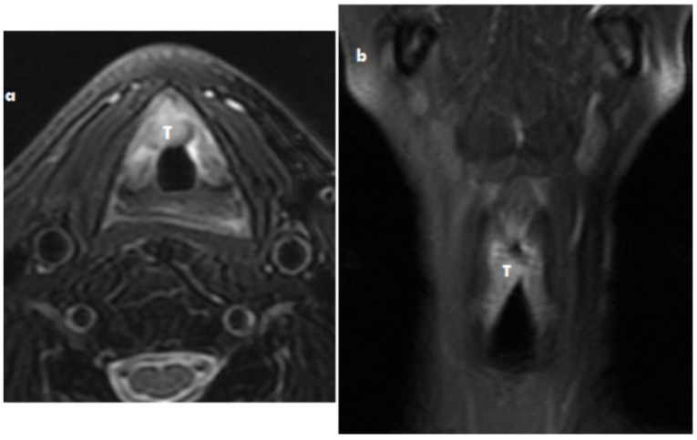
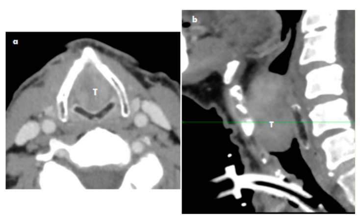
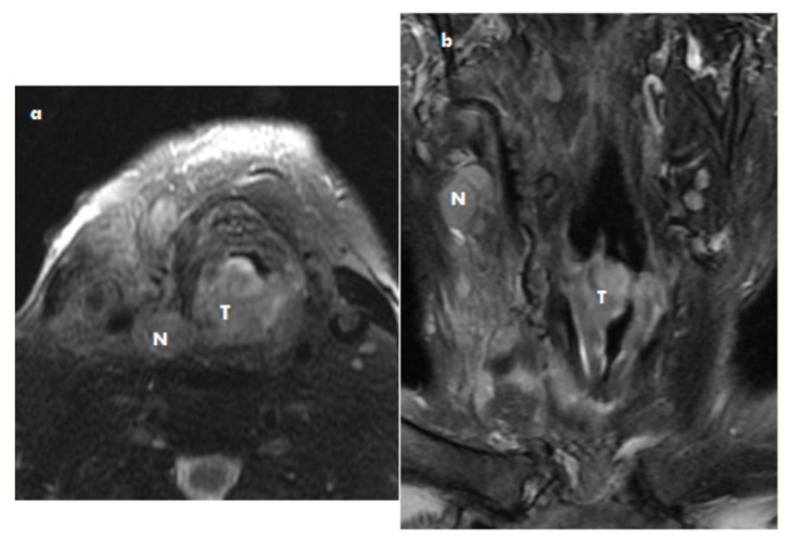
Soft tissue sarcomas in the head and neck are rare malignancies. They occur in this area in less than 1% of all malignant tumors. Some authors have described the development of sarcoma from the mesenchymal tissue in the larynx. The histological diagnosis of a sarcoma depends on the immunohistochemical investigation. In particularly difficult diagnoses, electron microscopy has to be used. The treatment recommendation depends on the histological type of sarcoma. We analysed and summarized data on the diagnostic criteria and therapy for sarcoma of the larynx presented in the literature. We present three new cases of laryngeal sarcoma and describe the analyses of the published diagnostic and treatment schedules of laryngeal sarcomas. We developed a treatment protocol recommendation for laryngeal sarcoma based on an analysis of literature data and case reports. This recommendation is based on histological type, staging, grading, size, and survival data.
Keywords: laryngectomy; laryngoscopy; larynx; sarcoma; surgery; surviving; treatment.
Conflict of interest statement
The authors declare no conflict of interest.
Figures
References
-
- Astl J., Synkova B., Kovar D., Rotnagl J., Tuckova I., Belsan T., Georgiev P. Leiomyosarcoma of the larynx—Diagnosis and Treatment protocol A case report and Review of the literature. Otolaryng Foniat. 2019;62:136–139.
Publication types
MeSH terms
Grants and funding
LinkOut - more resources
Full Text Sources
Other Literature Sources
Medical
