Review
. 2021 Feb 25;13(3):729.
doi: 10.3390/nu13030729.

Interactions between Growth of Muscle and Stature: Mechanisms Involved and Their Nutritional Sensitivity to Dietary Protein: The Protein-Stat Revisited

Affiliations
Review

Interactions between Growth of Muscle and Stature: Mechanisms Involved and Their Nutritional Sensitivity to Dietary Protein: The Protein-Stat Revisited

D Joe Millward. Nutrients. .

Abstract

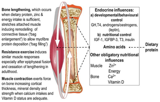
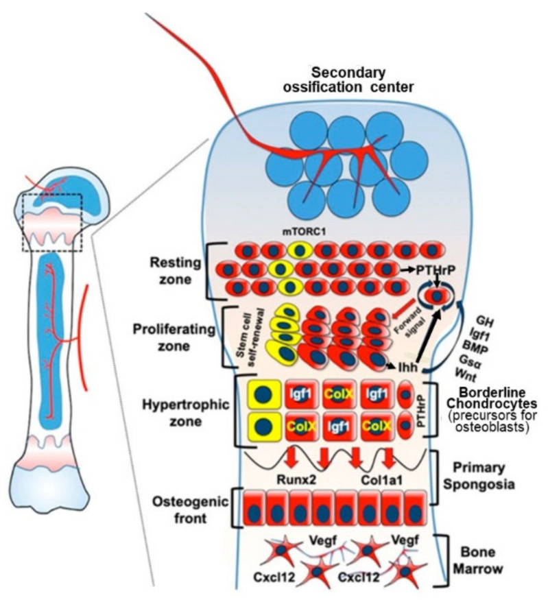
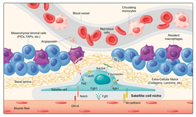
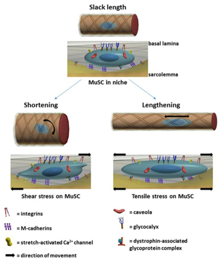
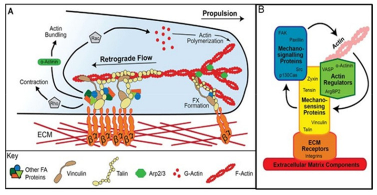
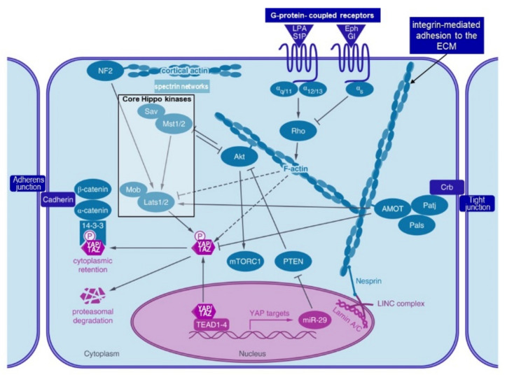
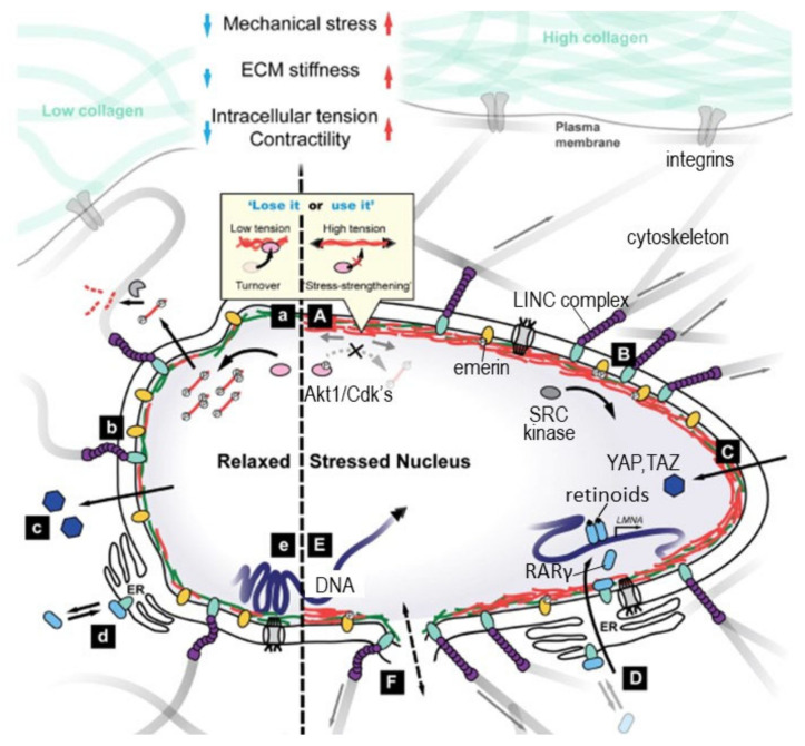
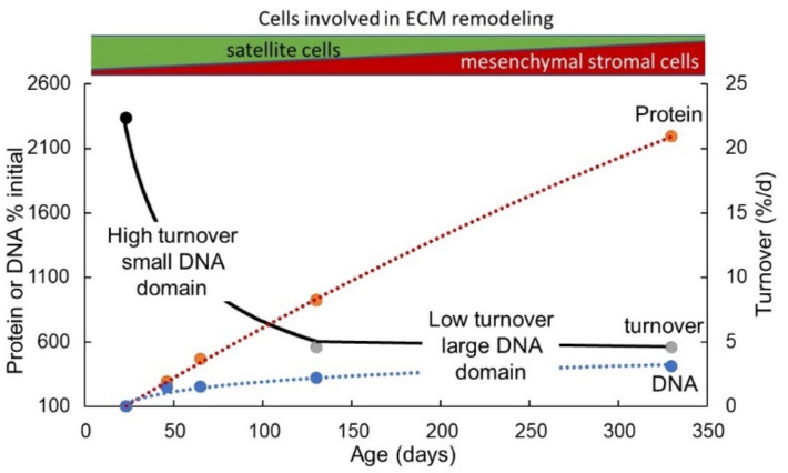
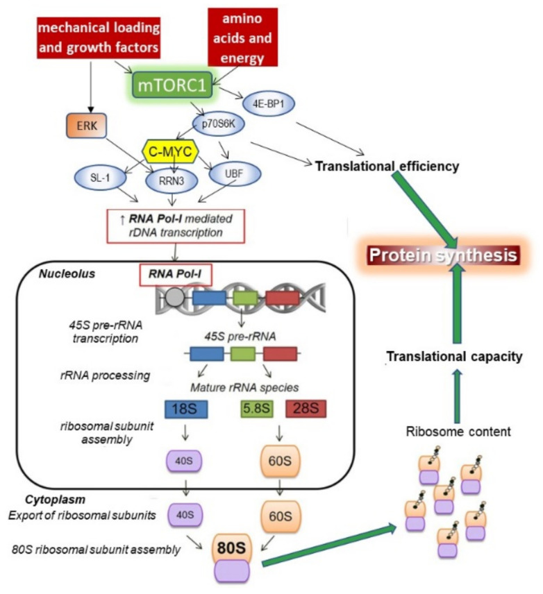
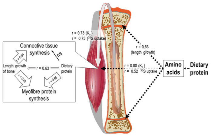
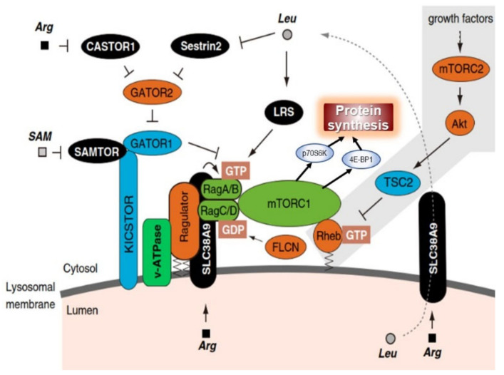
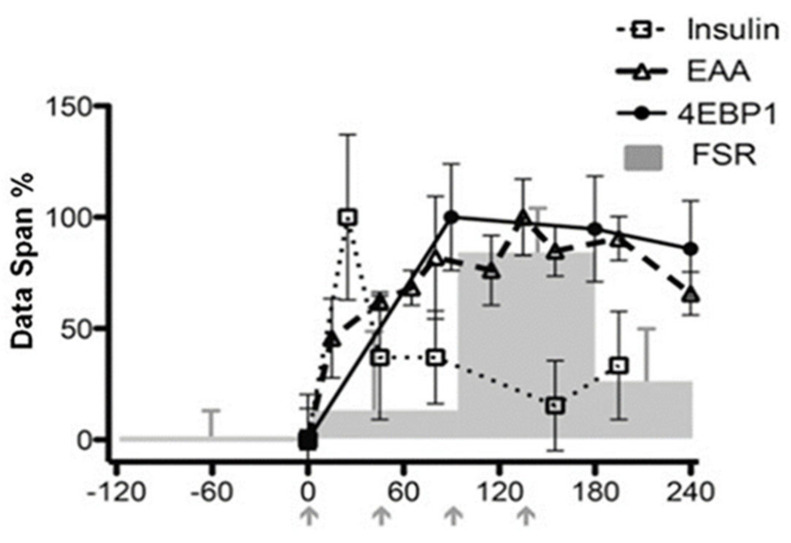
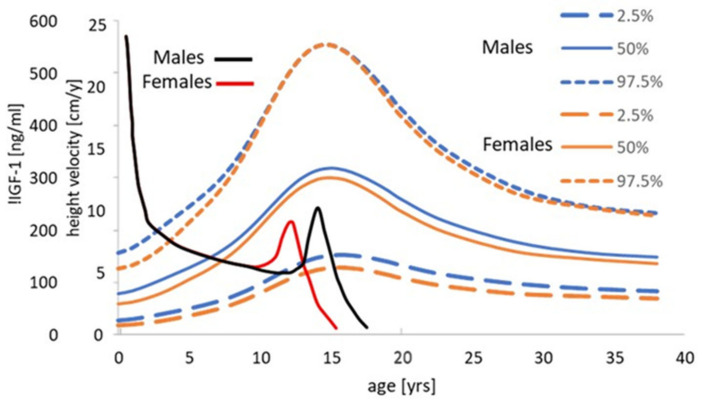
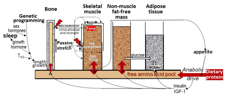
Childhood growth and its sensitivity to dietary protein is reviewed within a Protein-Stat model of growth regulation. The coordination of growth of muscle and stature is a combination of genetic programming, and of two-way mechanical interactions involving the mechanotransduction of muscle growth through stretching by bone length growth, the core Protein-Stat feature, and the strengthening of bone through muscle contraction via the mechanostat. Thus, growth in bone length is the initiating event and this is always observed. Endocrine and cellular mechanisms of growth in stature are reviewed in terms of the growth hormone-insulin like growth factor-1 (GH-IGF-1) and thyroid axes and the sex hormones, which together mediate endochondral ossification in the growth plate and bone lengthening. Cellular mechanisms of muscle growth during development are then reviewed identifying (a) the difficulties posed by the need to maintain its ultrastructure during myofibre hypertrophy within the extracellular matrix and the concept of muscle as concentric "bags" allowing growth to be conceived as bag enlargement and filling, (b) the cellular and molecular mechanisms involved in the mechanotransduction of satellite and mesenchymal stromal cells, to enable both connective tissue remodelling and provision of new myonuclei to aid myofibre hypertrophy and (c) the implications of myofibre hypertrophy for protein turnover within the myonuclear domain. Experimental data from rodent and avian animal models illustrate likely changes in DNA domain size and protein turnover during developmental and stretch-induced muscle growth and between different muscle fibre types. Growth of muscle in male rats during adulthood suggests that "bag enlargement" is achieved mainly through the action of mesenchymal stromal cells. Current understanding of the nutritional regulation of protein deposition in muscle, deriving from experimental studies in animals and human adults, is reviewed, identifying regulation by amino acids, insulin and myofibre volume changes acting to increase both ribosomal capacity and efficiency of muscle protein synthesis via the mechanistic target of rapamycin complex 1 (mTORC1) and the phenomenon of a "bag-full" inhibitory signal has been identified in human skeletal muscle. The final section deals with the nutritional sensitivity of growth of muscle and stature to dietary protein in children. Growth in length/height as a function of dietary protein intake is described in the context of the breastfed child as the normative growth model, and the "Early Protein Hypothesis" linking high protein intakes in infancy to later adiposity. The extensive paediatric studies on serum IGF-1 and child growth are reviewed but their clinical relevance is of limited value for understanding growth regulation; a role in energy metabolism and homeostasis, acting with insulin to mediate adiposity, is probably more important. Information on the influence of dietary protein on muscle mass per se as opposed to lean body mass is limited but suggests that increased protein intake in children is unable to promote muscle growth in excess of that linked to genotypic growth in length/height. One possible exception is milk protein intake, which cohort and cross-cultural studies suggest can increase height and associated muscle growth, although such effects have yet to be demonstrated by randomised controlled trials.

Keywords: IGF-1; amino acids; bone; dietary-protein; growth; insulin; mechanotransduction; muscle; protein-synthesis; satellite-cells.


Conflict of interest statement

The author declares no conflict of interest. The author has received consulting, travel, and/or speaker fees and research support (last 5 years) from Abbott, Nestlé Health Science, Public Health England and the UK Ministry of Defence.

Figures

Figure 1
Figure 1
Dietary protein and appendicular muscle- bone interactions.
Figure 2
Figure 2
Endochondral ossification: molecular characteristics of growth plate chondrocytes and their differentiation trajectories. Stem cells in the resting zone express the transcription factor SOX9 which drives their commitment and differentiation into chondrocytes. The initial development of the secondary ossification centre stimulates the expression of parathyroid hormone-related protein (PTHrP) by skeletal stem cells in the resting zone niche. This population contributes to the formation of proliferating and hypertrophic chondrocytes in addition to marking collagen Type I Alpha 1 Chain, (Col1a1+), osteoblasts in the primary spongiosa and stromal cell-derived factor 1, (Cxcl12), expressing bone marrow stromal cells in the marrow cavity over time. PTHrP acts to delay differentiation and keep the chondrocytes in a proliferative state, becoming organised into clones that arrange themselves into columns, contributing to bone elongation via interaction with adjacent cells. However, when these cells become sufficiently distant from the source of PTHrP due to interstitial growth, they differentiate into pre-hypertrophic and early hypertrophic chondrocytes, which are postmitotic cells that express Indian hedgehog (Ihh). This factor diffuses up the column to stimulate further PTHrP secretion to maintain the proliferative nature of the chondrocytes. After terminal differentiation, chondrocytes enlarge and this is the major contributing factor regulating the growth rate in endochondral bones. mTORC1 activity maintains the stem cells in their niche and remains important throughout proliferation and hypertrophy. Growth hormone acts within the resting zone to increase expression of IGF-1 and its receptor which is important for the PTHrP-Ihh feed-back loop and for the process of hypertrophy. T3 (not shown) is widely involved with indirect effects involving enhancing IGF-1 action and directly inhibiting proliferation and stimulating pre-hypertrophic and hypertrophic chondrocyte differentiation [120]. Bone morphogenetic proteins (BMPs) and their receptors are expressed in proliferating and hypertrophic chondrocytes and BMP signalling regulates chondrocyte proliferation and differentiation in part within the PTHrP-Ihh feed-back loop and by maintaining SOX9 activity. Gsα is a GTPase which activates adenyl cyclase to increase cyclic AMP and mediates the action of PTHrP and inhibits Ihh. Wnt(Wingless-related integration site signaling glycoproteins)/β-catenin signalling promotes differentiation of proliferating chondrocytes into hypertrophic chondrocytes both by antagonising PTHrP and independently of PTHrP signalling. Copied and adapted from Hallett et al. (2019) [48] under the Creative Commons Attribution 4.0 International license.
Figure 3
Figure 3
The three connective tissue layers or “bags” which define the volume of an individual skeletal muscle. The entire muscle, (top) is encased by the epimysium. Bundles of muscle fibres, (fascicles, middle), are encased by the perimysium. Each muscle cell, (middle and bottom), the multinucleate myofibre containing the myofibrillar contractile apparatus, is bounded by the sarcolemma or plasma membrane which is encased by the endomysium or basal lamina. Muscle stem cells (satellite cells) reside outside the myofibre between the plasma membrane and the basal lamina. Copied and adapted under the Creative Commons Attribution 4.0 International license from [132,133].
Figure 4
Figure 4
The satellite cell and stromal cell niche [139]. Satellite cells states are regulated through their interactions with their microenvironment. While direct interactions (M-cadherin, Notch pathway) and communication (FGF2/FGFR1 pathway) between muscle fibres and satellite cells have been identified, muscle stem cells also interact with a variety of components of the extracellular matrix (e.g., Collagens VI and V, Laminin, Fibronectin, SDC3/4) and diffusible cytokines and growth factors (e.g., Angiopoietin-Tie2 receptor). In addition to satellite cells, several cell types contribute to muscle growth, homoeostasis and regeneration, including pericytes, mesenchymal stromal cells (e.g., Pw1+ Interstitial Cells, Fibro Adipogenic Progenitors, Twist2+ cells) immune cells (e.g., resident or infiltrating macrophages) as well as connective tissue cells. Copied under the Creative Commons Attribution 4.0 International license from Evano and Tajbakhsh, 2018 [139].
Figure 5
Figure 5
Myogenic lineage progression and expression profile of key myogenic regulators [155]. Top Schematic illustration of the myogenic lineage progression. Satellite cells in their niche are activated by overstretch, start to proliferate, thereby generating myogenic progenitor cells. Upon differentiation, myogenic progenitor cells differentiate into myocytes, which fuse with the myofibre to form new myonuclei enabling myofibre hypertrophy and muscle growth. Middle Expression profile of key modulators of myogenic lineage progression. Sprouty 1 expression maintains a mitotically inactive state similar to quiescence [156]. The transcription factor Pax7 is required for the postnatal maintenance of satellite cells (SCs) and to enable their activation, but is lost after activation and proliferation and it cannot be identified in myonuclei after fusion with the myofibre [157,158]. Of the main myogenic regulatory factors (Myf5, MyoD, myogenin and Mrf4), MyoD is an early marker for myogenic commitment and Myogenin is a direct target of MyoD and initiates the terminal differentiation of myogenic progenitor cells, which is accompanied by downregulation of MyoD expression. The terminally differentiated myocytes fuse with the myofibre and express muscle genes such as myosin heavy chain (MHC). Bottom. A switch from Notch to Wnt signalling is required for proper satellite cell differentiation. Satellite cells express high levels of Notch to retain them in a quiescent state and upon activation canonical Wnt signalling increases. Canonical Wnt signalling—mainly through the ligand Wnt3a—drives differentiation of satellite cells, while non-canonical Wnt signalling through the ligand Wnt7a is responsible for promoting symmetric satellite cell divisions, migration of satellite cells, fusion with and growth of myofibres. Upon return to quiescence, satellite cells switch to Notch signalling. Copied and modified from Schmidt et al., 2019 [155] under the terms of the Creative Commons Attribution 4.0.
Figure 6
Figure 6
Potential effects of different types of stress on satellite cells. In response to mechanical overload by exercise or stretching, SCs are activated and proliferate to fuse with the host myofibre as shown in Figure 5. Both shear stress through shortening and tensile stress through lengthening may influence satellite cell orientation and deformation. In the unstrained (slack) myofibre is surrounded by a collagen fibre reinforced matrix (top). Below is shown an enlarged lateral view of the SC in its niche between the sarcolemma (yellow) and the basal lamina (BL). Transmembrane proteins anchoring the SC to the sarcolemma include M-cadherins and the dystrophin-associated glycoprotein complex. Both integrins and the glycocalyx anchor it to the BL. Caveolae are invaginations of the plasma membrane which function as message centres for regulating signal transduction and are identified by caveolin-1 which functionally regulates the activation state of caveolae-associated molecules, and which is associated with satellite cells in a more quiescent state [164]. With myofibre shortening, the sarcolemma will move relative to the BL and this is likely to twist the SC from a longitudinal orientation towards a more radial orientation (direction of arrow). As the myofibre is shortening, the sarcolemma and BL are likely moving at varying speeds, the relative movement of the sheaths will impose a shear force onto the SC, with subsequent cellular deformation. Myofibre lengthening will stretch both the sarcolemma and the BL (direction of arrow), which may induce a tensile stress on the SC. These physical cues, some acting through focal adhesion complexes, are thought to mediate the activation and differentiation of SC. Copied from Boers et al., 2018 [38] under the terms of the Creative Commons Attribution 4.0.
Figure 7
Figure 7
Integrin-based models of mechanotransduction which could operate in muscle satellite cells. (A) In the clutch model for SC migration, actin polymerisation at the advancing leading edge of the cell generates a retrograde flow of F-actin. Talin and vinculin both engage with the retrograde flow of actin, which generates tensional forces across them, regulating their activation states. Talin and vinculin bind to several signalling proteins including focal adhesion kinase, paxillin and others, producing downstream signalling events regulating the activity of the Rho-family GTPases. Vinculin can also induce actin bundling, acting through α-actinin. The activation state of talin and vinculin regulates their dynamic turnover, as well as influencing the molecular stoichiometry of the adhesion to direct downstream signalling to Rho-family GTPases. Rho activation leads to greater contractility and FA growth, whereas Rac activity enhances actin polymerisation at the leading edge. The growth of new actin filaments also leads to the formation of focal complexes (FXs), which can mature into larger focal adhesions, which act locally to control intracellular signalling. The sensing of mechanical stimulation (i.e., pulling on the integrin receptors during ECM stretching), or by the cell actively probing its ECM environment, utilises this linkage between the actin cytoskeleton and integrin-associated complexes (IACs) to transduce cellular migration. (B) IAC proteins can be roughly classified into the different groups shown based on their turnover. The ECM components (e.g., fibronectin) themselves are turned over, albeit slowly. The ECM receptors, such as integrins, have a slower turnover than any other IAC components. Mechanosensing proteins are those whose turnover rate has been reported to change in response to substrate stiffness. Mechanosignalling proteins do not change their turnover speed in response to substrate stiffness but are essential for generating the intracellular responses to mechanical stimuli. Finally, IACs contain proteins, the actin regulators with a slower turnover than the mechanosignalling proteins but faster than the mechanosensing proteins, which are responsible for locally regulating the actin cytoskeleton. Copied and modified from Jansen, Atherton and Ballestrem, 2017 [56] under the terms of the Creative Commons Attribution 4.0.
Figure 8
Figure 8
The hippo pathway and the role of YAP and TAZ in mechanotransduction of SC activity. The core components of the hippo pathway are serine/threonine kinases Mammalian Ste20-like 1/2 kinase (MST1/2) and large tumour suppressor 1/2 kinase (LATS1/2), which act to phosphorylate YAP (Yes-associated protein) and TAZ (transcriptional co-activator with PDZ binding motif), to determine the activation state of the pathway. When YAP and TAZ are unphosphorylated they are active and able to enter the nucleus and activate transcriptional enhancer factor, (TEAD)-mediated gene expression. After phosphorylation by LATS1/2 kinase, YAP binds to 14-3-3 proteins, leading to its cytoplasmic retention and also to its degradation. YAP activity is regulated by the actin cytoskeleton which inhibits YAP/TAZ phosphorylation and which is linked to the ECM via integrins. Actin stress fibres connect to the lamin meshwork in the nucleus via the LINC-complex, (Linker of Nucleoskeleton and Cytoskeleton). Rho GPTases are regulated by G-protein coupled receptor signalling which in turn regulates actin dynamics and YAP activity (dashed lines). Actin-binding proteins, like angiomotin (AMOT) or neurofibromin 2 (NF2/Merlin) are also known to regulate YAP activity, either through LATS or by direct interaction with YAP. Akt, a key regulator of the IGF-1-mTOR pathway, also binds to actin stress fibres and crosstalks to the Hippo pathway by interacting with MST1/2 and by YAP-induced expression of a microRNA (miR-29) which inhibits the inhibition of Akt by targeting the phosphatase and tensin homolog, PTEN. Copied and modified from Fischer et al. 2016 [231] with extra detail from [232].
Figure 9
Figure 9
Mechanosensing by the nucleus. (A and a) High nuclear tension can induce conformational changes in lamin A, C and B1/B2 coiled-coil dimers, which sterically inhibits access by kinases, Cyclin-dependent kinases (Cdk’s), protein kinase C (PKC), and protein kinase B, (Akt), which would otherwise increase lamin turnover. (B and b) Pulling on nesprin-1, (the LINC complex of Nesprin and sun domain-containing protein 2 (SUN2) which tethers the nucleus to the cytoskeleton), leads to phosphorylation of emerin by non-receptor tyrosine kinase (SRC) with stress stiffening of the nucleus influencing downstream mechanoresponses: e.g., formation of stress fibres, migration, localisation of YAP and TAZ, and serum response factor transcription. (C and c) Mechanosensitive transcription factors such as YAP and TAZ, which influence growth in the Hippo pathway, (see Figure 8), translocate into the nucleus under stress to modulate gene expression. (D and d) Mechanical stress leads to nuclear localisation of RARγ, (retinoic acid receptor γ) which directly regulates LMNA (lamins A and C gene) transcription. (E and e) Application of mechanical force may lead to changes in chromatin conformation (e.g., local stretching of genes), thereby altering transcriptional activity. (F) High tension can induce membrane dilation and may lead to transient ruptures, allowing for the exchange and mislocalisation of nucleoplasmic and cytoplasmic factors. Copied and modified from Cho, Irianto, and Discher 2017 [212] under a Creative Commons License (Attribution–Noncommercial–Share Alike 4.0 International license).
Figure 10
Figure 10
Myofibre growth and protein turnover in rat muscle. At weaning, the protein:DNA ratio is small partly because of a high proportion of satellite cells but also because within the myofibre the DNA domain size is small because each nucleus has to manage protein with a high turnover rate. The fall in the turnover rate with age allows each myonucleus to manage a larger domain allowing rapid growth of myofibre protein. The turnover rate shown is the fractional breakdown rate calculated as the measured in vivo rate of protein synthesis shown in well-fed rats in Table 2 minus the muscle growth rate [255].
Figure 11
Figure 11
Regulation of ribosomal capacity and efficiency of muscle protein synthesis by nutritional and mechanical factors. Multiple signalling pathways including extracellular signal-regulated kinase (ERK) and mTORC1 mediate activation of the nucleolar RNA Polymerase I (Pol-I) by a number of factors which form a transcriptional complex, the “Pol I regulon”, at the rDNA promoter [275]. The elevated expression of the proto-oncogene transcription factor C-MYC, achieved through mTORC1/S6K1 [287], plays a central role stimulating the Pol II-dependent transcription of a cohort of factors associated with Pol I regulon including RNA polymerase I-specific transcription initiation factor RRN3, (also known as TIF-1A, transcription initiation factor 1A) and upstream binding factor (UBF). Ribosome biogenesis involves transcription of the 45S ribosomal RNA precursor (45S pre-rRNA), processing of the 45S pre-rRNA into the smaller rRNAs (18S, 5.8S and 28S rRNAs) followed by assembly of these rRNAs and other ribosomal proteins into ribosomal subunits (40S and 60S) which are exported into the cytoplasm. The 80S ribosome is the mRNA translating unit, requiring mTORC1-mediated activation of initiation through p70S6K (p70 kDa ribosomal protein subunit kinase 1) and 4E-BP1 (eukaryotic initiation factor 4E binding protein 1). mTORC1 also regulates RNA Pol-II (which transcribes the ribosomal proteins and other proteins required for transcription) and Pol–III, (which synthesises small structural RNAs such as 5S rRNA and transfer RNA (tRNA) (not shown). Modified from Fyfe et al. 2018 [288] under a Creative Commons Attribution 4.0 International License.
Figure 12
Figure 12
Influences of dietary protein on muscle and bone growth and their interactions [291]. Relationships indicated by full and partial correlation analysis of the interaction between dietary protein intake and bone length growth as determinants of muscle myofibre protein synthesis (Ks) and muscle connective tissue synthesis (35S uptake) in the young rat measured during the first 3 days of variable dietary protein intakes (20%, 7%, 3.5% and 0.5% dietary protein). Bone length growth, muscle Ks and 35S-uptake were each significantly correlated with dietary protein intake and muscle Ks and 35S-uptake were each correlated with bone length growth. The independent influences of bone length growth and dietary protein on muscle is indicated by the partial r values in the box on the left, showing that the apparent influence of protein intake on 35S uptake in muscle is entirely accounted for by its influence on bone length growth, which in turn influences muscle 35S uptake and myofibre Ks to a lesser extent. Bone length growth was indicated by the more sensitive index of epiphysial cartilage width which is proportional to and an early indicator of, changes in overall tibial length [289].
Figure 13
Figure 13
Consensus model of amino acid-dependent mTORC1 activation at the lysosome [306]. Leucine (Leu), arginine (Arg), and S-adenosylmethionine (SAM) are sensed by cytosolic and lysosomal sensors, Sestrin2, CASTOR1, and SAMTOR, (S-adenosylmethionine sensor upstream of mTORC1), as well as LRS, (leucyl-tRNA synthetase 1) and SLC38A9, (essential amino acids transporter). These signals relieve inhibitory signalling and converge on Rag GTPases (heterodimeric RagA/B-RagC/D GTPases, members of a subfamily of the Ras-like small GTPase superfamily), which recruit mTORC1 onto the lysosomal surface. There mTORC1 is activated by association with the Rheb, (Ras homolog enriched in brain), GTPase. The efflux of leucine from the lysosomal lumen to the cytosol through the arginine-sensitive SLC38A9 transporter also activates mTORC1 via cytosolic sensors. Activated mTORC1 signals an increase in protein synthesis through p70S6K and 4E-BP1 (see [308] for more details of translational control). Copied and adapted from Takahara et al., 2020 [306] under the terms of the Creative Commons Attribution 4.0.
Figure 14
Figure 14
“Bag-full” effect in human skeletal muscle observed in the response of muscle protein synthesis to amino acid feeding [340]. Changes in muscle protein synthesis (FSR) measured in muscle biopsies at −2, 0, 1.5, 3 and 4 h, plasma essential amino acids (EAA), insulin concentrations and phosphor-4EBP1Thr65/70 (eukaryotic translation initiation factor 4E-binding protein 1) from fasted to fed normalised to their own data spans shown on the same axis in young men after consumption of 4 meals of 3.75 g of essential amino acids every 45 min as indicated by the grey arrows. Values are means ± SEMs, n = 8. The FSR was increased between 90 and 180 min (p < 0.05) but returned to baseline after this even though plasma EAA concentrations and muscle 4EBP1 phosphorylation remained elevated. Similar findings were reported by Atherton et al., 2010 [338] with an initial stimulation of the FSR 100 min after a single 48 g whey-protein bolus coinciding with an increase in intramuscular leucine which remained elevated at 200 min at which time the FSR had returned to baseline. Copied and adapted from Mitchell et al., 2015 [340] under the terms of the Creative Commons Attribution 3.0.
Figure 15
Figure 15
Serum IGF-1 and height velocity curves for males and females from birth to middle age [370]. It is clear that the overall pattern of changes in serum IGF-1 does not reflect height velocity apart from in puberty and even then, peak height velocity precedes peak IGF-1 by 2 years. Reference serum IGF1 percentiles for males and females calculated for a multicentre study with samples from 12 cohorts from the United States, Canada, and Europe (n = 15,014 subjects, 6697 males, 8317 females), with values obtained with an automated chemiluminescence IGF-I immunoassay. Values at birth from cord blood. Values drawn from Bidlingmaier et al. [370]. Height velocity curves for a cohort of Italian boys and girls [371].
Figure 16
Figure 16
The Protein-Stat, (updated from Millward 1995 [7]). Whole-body protein content is controlled through an amino-static appetite mechanism, acting primarily to maintain skeletal-muscle mass at a level set by the linear dimensions of the organism. Bone lengthening occurs at rates determined by genetic programming (canalisation) mediated by growth hormone, secretion of which is primarily sleep-dependent, by thyroid hormones and by the sex steroids during puberty, acting together with an appropriate anabolic drive deriving from dietary protein. This anabolic drive, both permissive and active, is mediated by amino acids, insulin, IGF-1 (and in the rat T3) and other important nutrients like zinc and vitamin D, and provides the regulatory stimulus for protein deposition in all tissues. Net protein deposition during skeletal muscle growth occurs within myofibres which are limited in volume by the extracellular matrix of connective tissue which surrounds individual and groups of myofibres as well as the whole muscle (see Figure 3), like concentric “bags”. Thus, increased myofibre diameter and length during muscle growth requires remodelling of the muscle ECM to enable “bag” enlargement which is mediated by the passive stretching of skeletal muscle subsequent to bone length growth, activating satellite cells and other associated cells through mechanotransduction mechanisms. The linkage of bone length to muscle mass allows muscle size to be regulated at a genotypic muscle weight–bone length ratio. Increasing muscle size and force generation acts via the mechanostat to increase bone mineralisation and strength commensurate with muscle mass. Any dietary protein-derived amino acid intake in excess of that required for maximal “bag filling” will either expand the non-muscle lean body mass or be oxidized with the carbon skeletons leaving the liver as ketones and glucose, the latter to be taken up in adipose tissue for lipogenesis. Also excess branched chain amino acids can be taken up directly by adipose tissue and converted to fat. This metabolic fate of excess protein as adipogenesis is part of the Early Protein Hypothesis in which excess protein intake in infancy programmes adiposity.

Similar articles

Cited by

References

    1. Tanner J. Growth as a measure of the nutritional and hygienic status of a population. Horm. Res. 1992;38:106–115. doi: 10.1159/000182580. - DOI - PubMed
    1. NCD Risk Factor Collaboration A century of trends in adult human height. Elife. 2016;5:1–29. doi: 10.7554/elife.13410. - DOI - PMC - PubMed
    1. Orsso C.E., Tibaes J.R.B., Oliveira C.L.P., Rubin D.A., Field C.J., Heymsfield S.B., Prado C.M., Haqq A.M. Low muscle mass and strength in pediatrics patients: Why should we care? Clin. Nutr. 2019;38:2002–2015. doi: 10.1016/j.clnu.2019.04.012. - DOI - PubMed
    1. Timpka S., Petersson I.F., Zhou C., Englund M. Muscle strength in adolescent men and risk of cardiovascular disease events and mortality in middle age: A prospective cohort study. BMC Med. 2014;12:1–8. doi: 10.1186/1741-7015-12-62. - DOI - PMC - PubMed
    1. Lee D.H., Keum N.N., Hu F.B., Orav E.J., Rimm E.B., Willett W.C., Giovannucci E.L. Predicted lean body mass, fat mass, and all cause and cause specific mortality in men: Prospective US cohort study. BMJ. 2018;362:k2575. doi: 10.1136/bmj.k2575. - DOI - PMC - PubMed

Substances

LinkOut - more resources