. 2021 Feb 25;11(3):606.
doi: 10.3390/ani11030606.

Evaluation of Triclosan Effects on Cultured Swine Luteal Cells

Affiliations

Evaluation of Triclosan Effects on Cultured Swine Luteal Cells

Giuseppina Basini et al. Animals (Basel). .

Abstract

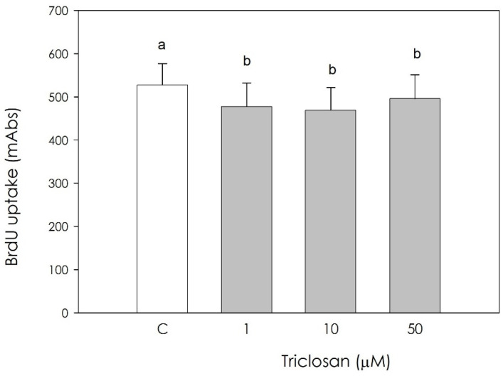
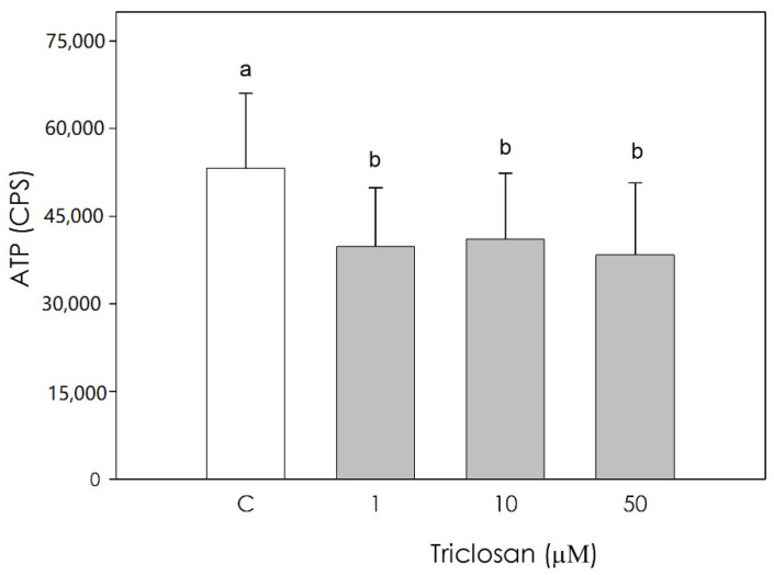
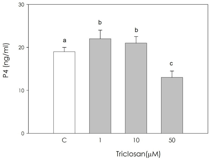
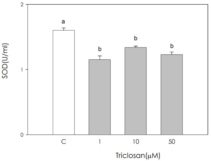
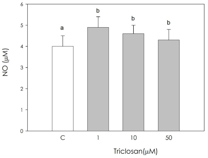
Triclosan is a chlorinated phenolic, used in many personal and home care products for its powerful antimicrobial effect. Several studies have shown triclosan toxicity and the American Food and Drug Administration (FDA) in 2016 has limited its use. It has been recently included in endocrine-disrupting chemicals (EDCs), a list of chemicals known for their ability to interfere with hormonal signaling with particular critical effects on reproduction both in animals and humans. In order to deepen the knowledge in this specific field, the present study was undertaken to explore the effect of different concentrations of triclosan (1, 10, and 50 µM) on cultured luteal cells, isolated from swine ovaries, evaluating effects on growth Bromodeoxyuridine (BrDU) incorporation and Adenosine TriPhosphate (ATP) production, steroidogenesis (progesterone secretion) and redox status (superoxide and nitric oxide production, enzymatic and non-enzymatic scavenging activity). A biphasic effect was exerted by triclosan on P4 production. In fact, the highest concentration inhibited, while the others stimulated P4 production (p < 0.05). Triclosan significantly inhibited cell proliferation, metabolic activity, and enzymatic scavenger activity (p < 0.05). On the contrary, nitric oxide production was significantly increased by triclosan (p < 0.01), while superoxide anion generation and non-enzymatic scavenging activity were unaffected.

Keywords: corpus luteum; nitric oxide; progesterone; redox status; superoxide anion.


Conflict of interest statement

The authors declare no conflict of interest.

Figures

Figure 1
Figure 1
Effect of the treatment with triclosan (1, 10, or 50 µM) for 48h on swine luteal cell proliferation using 5-bromo-2′-deoxyuridine (BrdU) incorporation assay test. Data, expressed as milliAbs units, represent the mean ± SEM of 36 replicates. Different letters on the bars indicate a significant difference (p < 0.05) among treatments as calculated by one-way ANOVA and Scheffè’ F test.
Figure 2
Figure 2
Effect of the treatment with triclosan (1, 10, or 50 µM) for 48 h on swine luteal cell metabolic activity using ATP assay test. Data, expressed as CPS, represent the mean ± SEM of 36 replicates. Different letters on the bars indicate a significant difference (p < 0.05) among treatments as calculated by one-way ANOVA and Scheffè’ F test.
Figure 3
Figure 3
Effect of the treatment with triclosan (1, 10, or 50 µM) for 48 h on swine luteal cell progesterone (P4) production using ELISA assay. Data, expressed as ng/mL, represent the mean ± SEM of 36 replicates. Different letters on the bars indicate a significant difference (p < 0.05) among treatments as calculated by one-way ANOVA and Scheffè’ F test.
Figure 4
Figure 4
Effect of the treatment with triclosan (1, 10, or 50 µM) for 48 h on swine luteal cell enzymatic scavenging activity using the SOD assay. Data, expressed as U/mL, represent the mean ± SEM of 36 replicates. Different letters on the bars indicate a significant difference (p < 0.05) among treatments as calculated by one-way ANOVA and Scheffè’ F test.
Figure 5
Figure 5
Effect of the treatment with triclosan (1, 10, or 50 µM) for 48 h on swine luteal cell nitric oxide (NO) production using Griess Assay. Data, expressed as µM, represent the mean ± SEM of 36 replicates. Different letters on the bars indicate a significant difference (p < 0.01) among treatments as calculated by one-way ANOVA and Scheffè’ F test.

Similar articles

Cited by

References

    1. Vosátka R., Krátký M., Vinšová J. Triclosan and its derivatives as antimycobacterial active agents. Eur. J. Pharm. Sci. 2018;114:318–331. doi: 10.1016/j.ejps.2017.12.013. - DOI - PubMed
    1. González-Casanova J.E., Pertuz-Cruz S.L., Caicedo-Ortega N.H., Rojas-Gomez D.M. Adipogenes is Regulation and Endocrine Disruptors: Emerging Insights in Obesity. Biomed. Res. Int. 2020;18:7453786. - PMC - PubMed
    1. Calafat A.M., Ye X., Wong L.Y., Reidy J.A., Needham L.L. Urinary concentrations of triclosan in the US population: 2003–2004. Environ. Health Perspect. 2008;116:303–307. doi: 10.1289/ehp.10768. - DOI - PMC - PubMed
    1. Wu J.L., Leung K.F., Sui-Fan T., Ching-Wan L. Organochlorine isotopic pattern-enhanced detection and quantification of triclosan and its metabolites in human serum by ultra-high-performance liquid chromatography/quadrupole time-of-flight/mass spectrometry. Rapid Commun. Mass Spectrom. 2012;26:123–132. doi: 10.1002/rcm.5303. - DOI - PubMed
    1. Dayan A.D. Risk assessment of triclosan [Irgasan] in human breast milk. Food Chem. Toxicol. 2007;45:125–129. doi: 10.1016/j.fct.2006.08.009. - DOI - PubMed

LinkOut - more resources