. 2021 Feb 25;22(5):2297.
doi: 10.3390/ijms22052297.

Spinal Excitatory Dynorphinergic Interneurons Contribute to Burn Injury-Induced Nociception Mediated by Phosphorylated Histone 3 at Serine 10 in Rodents

Affiliations

Spinal Excitatory Dynorphinergic Interneurons Contribute to Burn Injury-Induced Nociception Mediated by Phosphorylated Histone 3 at Serine 10 in Rodents

Angelika Varga et al. Int J Mol Sci. .

Abstract

The phosphorylation of serine 10 in histone 3 (p-S10H3) has recently been demonstrated to participate in spinal nociceptive processing. However, superficial dorsal horn (SDH) neurons involved in p-S10H3-mediated nociception have not been fully characterized. In the present work, we combined immunohistochemistry, in situ hybridization with the retrograde labeling of projection neurons to reveal the subset of dorsal horn neurons presenting an elevated level of p-S10H3 in response to noxious heat (60 °C), causing burn injury. Projection neurons only represented a small percentage (5%) of p-S10H3-positive cells, while the greater part of them belonged to excitatory SDH interneurons. The combined immunolabeling of p-S10H3 with markers of already established interneuronal classes of the SDH revealed that the largest subset of neurons with burn injury-induced p-S10H3 expression was dynorphin immunopositive in mice. Furthermore, the majority of p-S10H3-expressing dynorphinergic neurons proved to be excitatory, as they lacked Pax-2 and showed Lmx1b-immunopositivity. Thus, we showed that neurochemically heterogeneous SDH neurons exhibit the upregulation of p-S10H3 shortly after noxious heat-induced burn injury and consequential tissue damage, and that a dedicated subset of excitatory dynorphinergic neurons is likely a key player in the development of central sensitization via the p-S10H3 mediated pathway.

Keywords: burn injury; epigenetic modification; histone; neuropeptides; nociception; superficial dorsal horn neuron.


Conflict of interest statement

The authors declare no competing interest.

Figures

Figure 1
Figure 1
Burn injury-induced S10H3 phosphorylation is mainly located in the superficial dorsal horn (SDH) and displays unequal distribution within the spinal lumbar segments (L1–L6) in mice. (a) Innoxious stimulation (37 °C for 2 min) did not induce the phosphorylation of S10H3 in the SDH. (b) Burn injury (60 °C for 2 min) induced a robust phosphorylation of S10H3 in the ipsilateral SDH only. Both sections (a,b) were taken from L5 spinal segments. Dotted lines indicate the border between gray and white matter. (c) Burn injury-induced p-S10H3 expression shows unequal rostrocaudal distribution, with a peak expression in L6. Data presented as box plots are from 80-µm-thick sections taken from each segment (4–9 sections per segment from 3 animals). Dots indicate outlier values. * p < 0.05.
Figure 2
Figure 2
Neurons in the superficial dorsal horn exhibiting p-S10H3 immunoreactivity after burn injury show close appositions with CGRP-containing peptidergic afferents. (a1,a2) Immunostaining with antibodies against p-S10H3 (red), CGRP (blue) and Nissl (green). (a2) Asterisks indicate p-S10H3-positive neurons presenting close appositions with CGRP-containing peptidergic terminals. (b1,b2) Immunostaining with antibodies against p-S10H3 (red), isolectin B4 (IB4) (purple) and Nissl (green). (b2) None of the three p-S10H3-immunopositive neurons (empty arrowheads) have IB4 terminals in their vicinity. (c) Immunostaining with antibodies against p-S10H3 (red), CGRP (blue) and IB4 (purple). Dotted lines indicate the border between gray and white matter. D, dorsal; L, lateral. (d) Distribution of p-S10H3-immunoreactive nuclei within the CGRP- and IB4-positive bands and also in the deeper laminae of the SDH of rats and mice (n = 2 animals per species). The CGRP band contains the vast majority of p-S10H3 nuclei in both species.
Figure 3
Figure 3
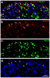
p-S10H3 was upregulated mainly but not exclusively in SDH neurons in mice after burn injury. (ad) Immunostaining with antibodies against p-S10H3 (red; b), Fox-3 (NeuN) (green; c), and Iba-1 (blue; d) in a transverse section of wild-type mouse. The merged image (a) clearly shows that almost all p-S10H3 positive nuclei are located in NeuN-positive somata (arrows) indicating that majority of them belong to neuronal population. Arrowheads label p-S10H3-expressing microglial cells. D, dorsal; L, lateral.
Figure 4
Figure 4
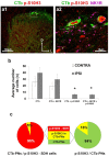
cholera toxin b-subunit (CTb)-labeled projection neurons rarely show nuclear burn injury-induced p-S10H3. (a1,a2) Immunostaining with antibodies against p-S10H3 (red), CTb (green) and neurokinin-1 receptor (NK1) receptor (NK1R; magenta) in an L5 transverse spinal cord section of a rat after burn injury. (a2) Higher magnification image represents the region with two CTb-labeled projection neurons (PNs) outlined by dashed line in (a1). These two CTb/p-S10H3 co-labeled neurons express NK1R. (b) Average number of CTb-labeled projection neurons (PNs) containing p-S10H3 positive nuclei and NK1R immunoreactivity (n = 17, 80-μm-thick sections per rat from 4 rats). IPSI refers to the side of the burn injury. Asterisks indicate a statistically significant difference between the ipsi- and contralateral sides of SDH (* p < 0.05). Note that the numbers of CTb-PNs with p-S10H3 or CTb/p-S10H3/NK1R ipsilaterally are negligible. Note also that the CTb-PNs could be detected on both sides. (c) Percentage of CTb-PNs within the total p-S10H3 positive SDH cell population (left) and percentage of PNs with p-S10H3 positive nuclei within the total CTb labeled PN population in the SDH (right).
Figure 5
Figure 5
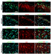
p-S10H3 is upregulated predominantly in excitatory interneurons both in wild-type and transgenic mice after burn injury. (ac) Immunostaining with antibodies against p-S10H3 (green) and Pax-2 (red) in a transverse section of a wild-type mouse. In this field of view, there is only one Pax-2-positive neuron that exhibits p-S10H3-IR in its nucleus (asterisk). Arrowheads mark p-S10H3-IR neurons which lack Pax-2. (df) Immunostaining with antibodies against p-S10H3 (green) and Lmx1b (red) in a transverse section of a wild-type mouse. In this field of view, there are numerous p-S10H3-positive neurons co-labeled with Lmx1b (arrowheads), while there is only one p-S10H3-IR cell that lacks Lmx1b (arrow). The dotted line indicates the border between gray and white matter (af). (gi) Immunostaining with antibodies against p-S10H3 (green) and red fluorescent protein (RFP) (red; against to tdTomato) in a transverse section of a vesicular gamma-amino butyric-acid (GABA) transporter (VGAT):tdTomato transgenic animal. There are three p-S10H3-positive neurons that exhibit weak RFP labeling in their cytoplasm (asterisks). Several p-S10H3-IR neurons without RFP-labeling can also be visible (arrowheads). (jl) Immunostaining with antibodies against p-S10H3 (green) and RFP (red; against to tdTomato) in a transverse section of a Vglut2:tdTomato transgenic animal. Nearly half of the p-S10H3-positive nuclei are located in tdTomato-positive somata (arrowheads), indicating their glutamatergic nature. There are additionally two p-S10H3-expressing neurons that lack RFP signals (marked with asterisks). D, dorsal; L, lateral.
Figure 6
Figure 6
Burn injury-induced S10H3 phosphorylation in excitatory subpopulations of SDH neurons. (a1a3) A representative image showing immunostaining for p-S10H3 (red; a1,a3) and somatostatin (SOM; green; a2,a3) in a transverse section of wild-type mouse. (a3) shows a merged image with 4′,6-diamidino-2-phenylindole (DAPI) (blue). In the inset there are two p-S10H3-positive neurons that exhibit strong SOM labeling in their cytoplasm (arrowheads). (b1b3) A representative image showing immunostaining for p-S10H3 (red; b1, b3) and substance P (SP; green; b2,b3) in a transverse section of wild-type mouse. (b3) shows a merged image with DAPI (blue). There are some SP-containing neuronal soma with (arrowheads) or without p-S10H3 (arrow) in their nuclei. Asterisks show p-S10H3-IR neurons that probably do not produce SP. (c1c5) A representative image showing immunostaining for p-S10H3 (red; c1,c3) and in situ hybridization (ISH) signal for Tac1 mRNA (green; c2,c3). (c3) shows a merged image with DAPI (blue). Insets represent higher magnification views of regions of interest designated on image (c3). Arrowheads label p-S10H3-IR neurons that probably do not produce substance P due to its low level of Tac1 mRNA (c4). Arrows indicates Tac1 mRNA containing cell bodies that do not show p-S10H3-IR (c5). (d) Immunostaining for p-S10H3 (green), cell nuclei specific DAPI (blue) and ISH signal for GRP mRNA (red). Interestingly, SDH neurons containing GRP mRNA (arrowheads) never express pS10H3-positive nuclei (asterisks) following burn injury. The dotted line indicates the border between gray and white matter (ad). D, dorsal; L, lateral; M, medial.
Figure 7
Figure 7
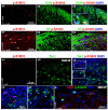
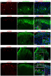
Burn injury-induced S10H3 phosphorylation in inhibitory subpopulations of SDH neurons. (a1a3) A representative image showing immunostaining for p-S10H3 (red; a1,a3), Nissl dye (blue; a3) and parvalbumin (green; a2a3). Nearly all p-S10H3-positive nuclei fell outside the band rich in parvalbumin-immunoreactive profiles and none was present in parvalbumin positive somata of deeper laminae (arrows). (b1b3) Immunostaining for p-S10H3 (red; b1,b3), Nissl dye (blue; b3) and calretinin (green; b2,b3). Calretinin positive neurons occasionally contained p-S10H3-positive nuclei (arrowhead in the inset). Asterisks indicate p-S10H3-IR neurons without calretinin immunolabeling. (c1c3) Immunostaining for p-S10H3 (red; c1,c3), neuronal marker, NeuN (blue; c3) and NPY (green; c2,c3). Hardly any of the rare NPY-IR neuronal somata contained a p-S10H3-positive nucleus as indicated with arrowheads. Asterisk marks p-S10H3-IR neurons without NPY immunolabeling. Arrow marks an NPY+ interneuron that lacks p-S10H3. (d1d3) Immunostaining with antibodies against p-S10H3 (red; d1,d3), NeuN (blue; d3) and nNOS (green; d2,d3). Asterisk indicates a p-S10H3-IR neuron without nNOS immunolabeling. Arrows show nNOS-IR neurons in which p-S10H3 was not upregulated upon burn injury. (e1e3) Immunostaining with antibodies against p-S10H3 (red; e1,e3), NeuN (blue; e3) and prodynorphin (Pdyn) (green; e2,e3). Several p-S10H3-immunostained nuclei were localized within dynorphin-positive somata (arrowheads in the inset). In the higher magnification image, an asterisk labels a non-dynorphinergic neuron that exhibits p-S10H3. Dotted lines indicate the border between gray and white matter (b3,c3,d3,e3).
Figure 8
Figure 8
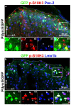
Burn injury-induced p-S10H3 nuclei are dominantly expressed by the excitatory subpopulation of dynorphinergic SDH neurons. (a) A representative image showing immunostaining for p-S10H3 (red), GFP (green) and Pax-2 (blue) in a transverse spinal cord section of Pdyn:enhanced green fluorescent protein (EGFP) mouse. In the inset (a1a4) there are several anti-GFP antibody-labeled dynorphinergic neurons. Of these, there is only one p-S10H3-expressing Pdyn+ neuron that exhibits Pax-2-IR (labeled with an arrow), while the others lack Pax-2 (arrowheads; a1a4). (b) A representative image showing immunostaining for p-S10H3 (red), GFP (green) and Lmx1b (blue) in a transverse spinal cord section of Pdyn:EGFP mouse. In the insets (b1b4), numerous p-S10H3-immunoreactive nuclei are visible in laminae I–IIo of the SDH. Of these, the majority express Lmx1b and thus presumably they are excitatory. While there are several p-S10H3+/Lmx1b+ neurons that proved to be dynorphinergic based on their strong cytoplasmic GFP-staining (arrowheads in the insets), there is only one that is non-dynorphinergic (indicated by an arrow). The asterisk indicates such a p-S10H3-IR neuron that is immunonegative both for Lmx1b and Pdyn. The dotted line represents the border between gray and white matter (a,b). D, dorsal; L, lateral. Scale bars in the insets are 20 µm.

References

    1. Sahbaie P., Liang D.Y., Shi X.Y., Sun Y., Clark J.D. Epigenetic regulation of spinal cord gene expression contributes to enhanced postoperative pain and analgesic tolerance subsequent to continuous opioid exposure. Mol. Pain. 2016;12:1744806916641950. doi: 10.1177/1744806916641950. - DOI - PMC - PubMed
    1. Khangura R.K., Bali A., Jaggi A.S., Singh N. Histone acetylation and histone deacetylation in neuropathic pain: An unresolved puzzle? Eur. J. Pharmacol. 2017;795:36–42. doi: 10.1016/j.ejphar.2016.12.001. - DOI - PubMed
    1. Tochiki K.K., Maiaru M., Norris C., Hunt S.P., Geranton S.M. The mitogen and stress-activated protein kinase 1 regulates the rapid epigenetic tagging of dorsal horn neurons and nocifensive behaviour. Pain. 2016;157:2594–2604. doi: 10.1097/j.pain.0000000000000679. - DOI - PMC - PubMed
    1. Torres-Perez J.V., Santha P., Varga A., Szucs P., Sousa-Valente J., Gaal B., Sivado M., Andreou A.P., Beattie S., Nagy B., et al. Phosphorylated Histone 3 at Serine 10 Identifies Activated Spinal Neurons and Contributes to the Development of Tissue Injury-Associated Pain. Sci. Rep. 2017;7:41221. doi: 10.1038/srep41221. - DOI - PMC - PubMed
    1. Todd A.J. Identifying functional populations among the interneurons in laminae I-III of the spinal dorsal horn. Mol. Pain. 2017;13:1744806917693003. doi: 10.1177/1744806917693003. - DOI - PMC - PubMed

MeSH terms

LinkOut - more resources