Review
. 2021 Feb 25;26(5):1232.
doi: 10.3390/molecules26051232.

Propolis, Bee Honey, and Their Components Protect against Coronavirus Disease 2019 (COVID-19): A Review of In Silico, In Vitro, and Clinical Studies

Affiliations
Review

Propolis, Bee Honey, and Their Components Protect against Coronavirus Disease 2019 (COVID-19): A Review of In Silico, In Vitro, and Clinical Studies

Amira Mohammed Ali et al. Molecules. .

Abstract

Despite the virulence and high fatality of coronavirus disease 2019 (COVID-19), no specific antiviral treatment exists until the current moment. Natural agents with immune-promoting potentials such as bee products are being explored as possible treatments. Bee honey and propolis are rich in bioactive compounds that express strong antimicrobial, bactericidal, antiviral, anti-inflammatory, immunomodulatory, and antioxidant activities. This review examined the literature for the anti-COVID-19 effects of bee honey and propolis, with the aim of optimizing the use of these handy products as prophylactic or adjuvant treatments for people infected with severe acute respiratory syndrome-coronavirus-2 (SARS-CoV-2). Molecular simulations show that flavonoids in propolis and honey (e.g., rutin, naringin, caffeic acid phenyl ester, luteolin, and artepillin C) may inhibit viral spike fusion in host cells, viral-host interactions that trigger the cytokine storm, and viral replication. Similar to the potent antiviral drug remdesivir, rutin, propolis ethanolic extract, and propolis liposomes inhibited non-structural proteins of SARS-CoV-2 in vitro, and these compounds along with naringin inhibited SARS-CoV-2 infection in Vero E6 cells. Propolis extracts delivered by nanocarriers exhibit better antiviral effects against SARS-CoV-2 than ethanolic extracts. In line, hospitalized COVID-19 patients receiving green Brazilian propolis or a combination of honey and Nigella sativa exhibited earlier viral clearance, symptom recovery, discharge from the hospital as well as less mortality than counterparts receiving standard care alone. Thus, the use of bee products as an adjuvant treatment for COVID-19 may produce beneficial effects. Implications for treatment outcomes and issues to be considered in future studies are discussed.

Keywords: ACE-II; COVID -19; SARS-CoV-2; bee honey; bee products; coronavirus disease 2019; coronaviruses; cytokine storm; flavonoids; in silico; in vitro; main protease; molecular docking/biochemical modeling; non-structural proteins; propolis; randomized clinical trials; severe acute respiratory syndrome; spike glycoprotein.


Conflict of interest statement

The authors declare no conflict of interest.

Figures

Figure 1
Figure 1
Schematic illustration of molecular modeling of interactions involving bioactive compounds in propolis and bee honey with SARS-CoV-2 proteins and host-cell receptor/proteases. 2D: two-dimensional; SARS-CoV-2: severe acute respiratory syndrome-coronavirus-2; 3CLpro: 3-chymotrypsin-like cysteine protease; RdRp: RNA-dependent RNA polymerase; PLpro: papain-like protease, ACE-II: angiotensin-converting enzyme-related carboxypeptidase; PP2A-B56: B56 regulatory unit of protein phosphatase 2 A; RCSBPDB: Research Collaboratory for Structural Bioinformatics Protein Data Bank; SLiMs: short linear motifs; ELM: eukaryotic linear motif resource. The two-dimensional (2D) or 3D structure of bee-derived compounds were obtained from ZINC database [25] (http://zinc15.docking.org/, accessed on 4 January 2021) or PubChem database (https://www.ebi.ac.uk/chembl/, accessed on 4 January 2021) [2,5,47,48]. Most studies retrieved the 3D crystal structures of ACE-II (PDB ID: R4L, the inhibitory bound state of the extracellular metallopeptidase domain of ACE-II with MLN-4760) [48], S1 subunit (PDB ID: 7BZ5) [51], PP2A-B56 (PDB ID: 5SWF-A) [2], and SARS-CoV-2 proteins such as S protein (PDB ID: 6m0j) [78] and (PDB ID: 7BZ5) [51], 3CLpro (PDB ID: 6LU7) [51], 3CLpro (PDB ID: 6Y2F, bound to α-ketoamide) [42], 3CLpro (PDB ID: 6W63, bound to ligand X77), Mpro (PDB ID: 5R7Y) [25], and RdRp (PDB ID:6M71) [25] from Research Collaboratory for Structural Bioinformatics Protein Data Bank (RCSBPDB) (http://www.rcsb.org, accessed on 4 January 2021) as PDB files. SLiMs (SARS-CoV-2 spike protein sequence) were scanned with the eukaryotic linear motif (ELM) resource (http://elm.eu.org/, accessed on 4 January 2021) [2]. Because the structure of TMPRSS2 was not available in RCSBPDB, one study modeled the structure of TMPRSS2 to a similar protease (human hepsin, obtained from the Swiss model repository (O15393)). The sequence of TMPRSS2 is 33.82% identical to hepsin; the catalytic domain in both proteins is well conserved with identical catalytic residues His296, Asp345, and Ser441, while the Q mean of the modeled structure was -1.62 [5]. Human cell enzymes (e.g., ACE-II and TMPRSS2) and SARS-CoV-2 proteins were geometrically optimized to act as receptors for virtual binding with bee-related compounds, which were also optimized to account for ligands in virtual binding models. The quality of docking was validated, and energy minimization was conducted [2].
Figure 2
Figure 2
Chemical structure of flavonoids with the highest binding affinity to target proteins of severe acute respiratory syndrome-coronavirus-2 (SARS-CoV-2).
Figure 3
Figure 3
3D structure of luteolin (a) and binding poses involving its interaction within the active sites of ACE-II (b), Mpro/3CLpro (c), and PLpro (d). Modified with permission from Shawan et al. [86], Bulletin of the National Research Centre, Springer Open, 2021, http://creativecommons.org/licenses/by/4.0/.
Figure 4
Figure 4
Binding pose (a) along with 2D (b) and 3D (c) analysis of molecular interaction of rutin within the active site of ACE-II. Reproduced with permission from Güler et al. [48], ScienceOpen Preprints, 2020, http://creativecommons.org/licenses/by/4.0/.
Figure 5
Figure 5
Binding pose (a) along with 2D (b) and 3D (c) analysis of molecular interaction of naringin within the active site of S protein. Reproduced with permission from Jain et al. [78], Saudi Journal of Biological Sciences, Elsevier, 2021, https://creativecommons.org/licenses/by-nc-nd/4.0/.
Figure 6
Figure 6
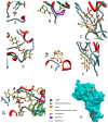
3D-plots for docking (A) Avigan, (B) hydroxychloroquine, (C) rutin, (D) caffeic acid phenethyl ester, (E) pinobanksin, (F) quercetin, and (G) remdesivir in the active site of S1 subunit of S protein, (PDB ID: 7BZ5). (H) 3D-plot comparing pose docking of Avigan and hydroxychloroquine to that of rutin, caffeic acid phenethyl ester, pinobanksin, and quercetin. Reproduced with permission from Refaat et al. [51], International Journal of Pharmaceutics, Elsevier B.V., 2020, https://creativecommons.org/licenses/by-nc-nd/4.0/.

References

    1. Goyal B., Goyal D. Targeting the Dimerization of the Main Protease of Coronaviruses: A Potential Broad-Spectrum Therapeutic Strategy. ACS Comb. Sci. 2020;22:297–305. doi: 10.1021/acscombsci.0c00058. - DOI - PubMed
    1. Maaroufi H. The Spike Protein S1 Subunit of SARS-CoV-2 Contains an LxxIxE-like Motif that is Known to Recruit the Host PP2A-B56 Phosphatase. bioRxiv. 2020:020941. doi: 10.1101/2020.04.01.020941. - DOI
    1. Zhou Y., Fu B., Zheng X., Wang D., Zhao C., Qi Y., Sun R., Tian Z., Xu X., Wei H. Pathogenic T-cells and inflammatory monocytes incite inflammatory storms in severe COVID-19 patients. Natl. Sci. Rev. 2020;7:998–1002. doi: 10.1093/nsr/nwaa041. - DOI - PMC - PubMed
    1. Ali A.M., Kunugi H. Corona Virus Disease 2019 (COVID-19): A pandemic that threatens physical and mental health by promoting physical inactivity. Sports Med. Health Sci. 2020;2:221–223. doi: 10.1016/j.smhs.2020.11.006. - DOI - PMC - PubMed
    1. Kumar V., Dhanjal J.K., Bhargava P., Kaul A., Wang J., Zhang H., Kaul S.C., Wadhwa R., Sundar D. Withanone and withaferin-A are predicted to interact with transmembrane protease serine 2 (TMPRSS2) and block entry of SARS-CoV-2 into cells. J. Biomol. Struct. Dyn. 2020:1–13. doi: 10.1080/07391102.2020.1775704. - DOI - PMC - PubMed